30
views
0
recommends
+1 Recommend
0 collections
    0
    shares
      • Record: found
      • Abstract: found
      • Article: found
      Is Open Access

      rTMS over bilateral inferior parietal cortex induces decrement of spatial sustained attention

      research-article

      Read this article at

      Bookmark
          There is no author summary for this article yet. Authors can add summaries to their articles on ScienceOpen to make them more accessible to a non-specialist audience.

          Abstract

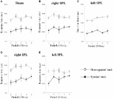
          Sustained attention is an essential brain function that enables a subject to maintain attention level over the time of a task. In previous work, the right inferior parietal lobe (IPL) has been reported as one of the main brain regions related to sustained attention, however, the right lateralization of vigilance/sustained attention is unclear because information about the network for sustained attention is traditionally provided by neglect patients who typically have right brain damage. Here, we investigated sustained attention by applying a virtual lesion technique, transcranial magnetic stimulation (TMS), over the left and right superior parietal lobe (SPL) and IPL. We used two different types of visual sustained attention tasks: spatial (location based) and non-spatial (feature based). When the participants performed the spatial task, repetitive TMS (rTMS) over either the right or left IPL induced a significant decrement of sustained attention causing a progressive increment of errors and response time. In contrast, participants' performance was not changed by rTMS on the non-spatial task. Also, omission errors (true negative) gradually increased with time on right and left IPL rTMS conditions, while commission errors (false positive) were relatively stable. These findings suggest that the maintenance of attention, especially in tasks regarding spatial location, is not uniquely lateralized to the right IPL, but may also involve participation of the left IPL.

          Related collections

          Most cited references35

          • Record: found
          • Abstract: found
          • Article: not found

          Responses to rapid-rate transcranial magnetic stimulation of the human motor cortex.

          We applied trains of focal, rapid-rate transcranial magnetic stimulation (rTMS) to the motor cortex of 14 healthy volunteers with recording of the EMG from the contralateral abductor pollicis brevis, extensor carpi radialis, biceps brachii and deltoid muscles. Modulation of the amplitude of motor evoked potentials (MEPs) produced in the target muscle during rTMS showed a pattern of inhibitory and excitatory effects which depended on the rTMS frequency and intensity. With the magnetic coil situated over the optimal scalp position for activating the abductor pollicis brevis, rTMS led to spread of excitation, as evident from the induction of progressively larger MEPs in the other muscles. The number of pulses inducing this spread of excitation decreased with increasing rTMS frequency and intensity. Latency of the MEPs produced in the other muscles during the spread of excitation was significantly longer than that produced by single-pulse TMS applied to the optimal scalp positions for their activation. The difference in MEP latency could be explained by a delay in intracortical conduction along myelinated cortico-cortical pathways. Following rTMS, a 3-4 min period of increased excitability was demonstrated by an increase in the amplitude of MEPs produced in the target muscles by single-pulse TMS. Nevertheless, repeated rTMS trains applied 1 min apart led to similar modulation of the responses and to spread of excitation after approximately the same number of pulses. This suggests that the spread might be due to the breakdown of inhibitory connections or the recruitment of excitatory pathways, whereas the post-stimulation facilitation may be due to a transient increase in the efficacy of excitatory synapses.
            Bookmark
            • Record: found
            • Abstract: found
            • Article: not found

            Vigilance requires hard mental work and is stressful.

            We describe major discoveries and developments in vigilance research. Vigilance tasks have typically been viewed as undemanding assignments requiring little mental effort. The vigilance decrement function has also been considered to result from a decline in arousal brought about by understimulation. Recent research in vigilance is reviewed in four areas: studies of task type, perceived mental workload during vigilance, neural measures of resource demand in vigilance, and studies of task-induced stress. Experiments comparing successive and simultaneous vigilance tasks support an attentional resource theory of vigilance. Subjective reports also show that the workload of vigilance is high and sensitive to factors that increase processing demands. Neuroimaging studies using transcranial Doppler sonography provide strong, independent evidence for resource changes linked to performance decrement in vigilance tasks. Finally, physiological and subjective reports confirm that vigilance tasks reduce task engagement and increase distress and that these changes rise with increased task difficulty. Converging evidence using behavioral, neural, and subjective measures shows that vigilance requires hard mental work and is stressful. This research applies to most human-machine systems that require human monitoring, particularly those involving automated subsystems.
              Bookmark
              • Record: found
              • Abstract: found
              • Article: not found

              Modulation of corticospinal excitability by repetitive transcranial magnetic stimulation.

              Repetitive transcranial magnetic stimulation (rTMS) is able to modulate the corticospinal excitability and the effects appear to last beyond the duration of the rTMS itself. Different studies, employing different rTMS parameters, report different modulation of corticospinal excitability ranging from inhibition to facilitation. Intraindividual variability of these effects and their reproducibility are unclear. We examined the modulatory effects of rTMS to the motor cortex at various frequencies (1, 10, 20 Hz) and at different time-points in twenty healthy volunteers. We observed significant inhibition of MEPs following 1 Hz rTMS and significant facilitation of MEPs following 20 Hz rTMS for both day1 and day 2. Interestingly, at 1 Hz and 20 Hz rTMS, the modulatory effect produced by rTMS was greater on day 2. However, there was no significant change in corticospinal excitability following 10 Hz rTMS neither on day 1 nor day 2. Our findings raise questions as to how stimulation parameters should be determined when conducting studies applying rTMS on multiple days, and in particular, studies exploring rTMS as a treatment modality in neuropsychiatric disorders.
                Bookmark

                Author and article information

                Journal
                Front Hum Neurosci
                Front Hum Neurosci
                Front. Hum. Neurosci.
                Frontiers in Human Neuroscience
                Frontiers Media S.A.
                1662-5161
                11 February 2013
                2013
                : 7
                : 26
                Affiliations
                [1] 1Department of Biomedical Engineering, Hanyang University Seoul, South Korea
                [2] 2Department of Biomedical Engineering, Keimyung University Daegu, South Korea
                [3] 3Department of Rehabilitation and Assistive Technology, National Rehabilitation Center Research Institute, Yonsei University College of Medicine Seoul, South Korea
                [4] 4Department of Neuropsychiatry and Clinical Research Institute, Seoul National University Hospital Seoul, South Korea
                [5] 5Department of Neurosurgery, MEG Center, Seoul National University College of Medicine Seoul, South Korea
                [6] 6Department of Psychiatry, Yonsei University College of Medicine Seoul, South Korea
                [7] 7Institute of Neurology and Institute of Cognitive Neuroscience, University College London London, UK
                [8] 8St. Peter's Hospital Seoul, South Korea
                [9] 9Osong Medical Innovation Foundation, Medical Device Development Center Chungbuk, South Korea
                Author notes

                Edited by: John J. Foxe, Albert Einstein College of Medicine, USA

                Reviewed by: Vincenzo Romei, University College London, UK; Mario Bonato, University of Padova, Italy

                *Correspondence: Dong Pyo Jang and Sun I. Kim, Department of Biomedical Engineering, Hanyang University, PO Box 17 Hangdang-dong, Seongdong-gu, Seoul 133-791, South Korea. e-mail: dongpjang@ 123456gmail.com ; sunkim7114@ 123456gmail.com
                Article
                10.3389/fnhum.2013.00026
                3568694
                23403477
                4e077f51-5507-4d09-91f0-6a92f9af8c80
                Copyright © 2013 Lee, Ku, Han, Park, Lee, Kim, Lee, Husain, Yoon, Kim, Jang and Kim.

                This is an open-access article distributed under the terms of the Creative Commons Attribution License, which permits use, distribution and reproduction in other forums, provided the original authors and source are credited and subject to any copyright notices concerning any third-party graphics etc.

                History
                : 27 August 2012
                : 21 January 2013
                Page count
                Figures: 4, Tables: 1, Equations: 0, References: 45, Pages: 8, Words: 5770
                Categories
                Neuroscience
                Original Research Article

                Neurosciences
                sustained attention,vigilance,repeated transcranial magnetic stimulation,inferior parietal lobe,spatial attention

                Comments

                Comment on this article