18
views
0
recommends
+1 Recommend
0 collections
    0
    shares
      • Record: found
      • Abstract: found
      • Article: found
      Is Open Access

      Galectin‐3, a Biomarker Linking Oxidative Stress and Inflammation With the Clinical Outcomes of Patients With Atherothrombosis

      research-article

      Read this article at

      Bookmark
          There is no author summary for this article yet. Authors can add summaries to their articles on ScienceOpen to make them more accessible to a non-specialist audience.

          Abstract

          Background

          Galectin‐3 (Gal‐3) participates in different mechanisms involved in atherothrombosis, such as inflammation, proliferation, or macrophage chemotaxis. Thus, there have been committed intensive efforts to elucidate the function of Gal‐3 in cardiovascular (CV) diseases. The role of Gal‐3 as a circulating biomarker has been demonstrated in patients with heart failure, but its importance as a biomarker in atherothrombosis is still unknown.

          Methods and Results

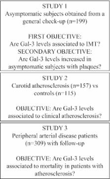
          Because Gal‐3 is involved in monocyte‐to‐macrophage transition, we used fresh isolated monocytes and the in vitro model of macrophage differentiation of THP‐1 cells stimulated with phorbol myristate acetate (PMA). Gal‐3 release is increased by PMA in human monocytes and macrophages, a process involving exosomes and regulated by reactive oxygen species/NADPH oxidase activity. In asymptomatic subjects (n=199), Gal‐3 plasma levels are correlated with NADPH oxidase activity in peripheral blood mononuclear cells ( r=0.476; P<0.001) and carotid intima‐media thickness ( r=0.438; P<0.001), a surrogate marker of atherosclerosis. Accordingly, Gal‐3 plasma concentrations are increased in patients with carotid atherosclerosis (n=158), compared to control subjects (n=115; 14.3 [10.7 to 16.9] vs. 10.4 [8.6 to 12.5] ng/mL; P<0.001). Finally, on a 5‐year follow‐up study in patients with peripheral artery disease, Gal‐3 concentrations are significantly and independently associated with an increased risk for CV mortality (hazard ratio=2.24, 95% confidence interval: 1.06 to 4.73, P<0.05).

          Conclusions

          Gal‐3 extracellular levels could reflect key underlying mechanisms involved in atherosclerosis etiology, development, and plaque rupture, such as inflammation, infiltration of circulating cells and oxidative stress. Moreover, circulating Gal‐3 concentrations are associated with clinical outcomes in patients with atherothrombosis.

          Related collections

          Most cited references24

          • Record: found
          • Abstract: found
          • Article: not found

          Proteomic analysis of dendritic cell-derived exosomes: a secreted subcellular compartment distinct from apoptotic vesicles.

          Dendritic cells constitutively secrete a population of small (50-90 nm diameter) Ag-presenting vesicles called exosomes. When sensitized with tumor antigenic peptides, dendritic cells produce exosomes, which stimulate anti-tumor immune responses and the rejection of established tumors in mice. Using a systematic proteomic approach, we establish the first extensive protein map of a particular exosome population; 21 new exosomal proteins were thus identified. Most proteins present in exosomes are related to endocytic compartments. New exosomal residents include cytosolic proteins most likely involved in exosome biogenesis and function, mainly cytoskeleton-related (cofilin, profilin I, and elongation factor 1alpha) and intracellular membrane transport and signaling factors (such as several annexins, rab 7 and 11, rap1B, and syntenin). Importantly, we also identified a novel category of exosomal proteins related to apoptosis: thioredoxin peroxidase II, Alix, 14-3-3, and galectin-3. These findings led us to analyze possible structural relationships between exosomes and microvesicles released by apoptotic cells. We show that although they both represent secreted populations of membrane vesicles relevant to immune responses, exosomes and apoptotic vesicles are biochemically and morphologically distinct. Therefore, in addition to cytokines, dendritic cells produce a specific population of membrane vesicles, exosomes, with unique molecular composition and strong immunostimulating properties.
            Bookmark
            • Record: found
            • Abstract: found
            • Article: not found

            Galectin-3 marks activated macrophages in failure-prone hypertrophied hearts and contributes to cardiac dysfunction.

            Inflammatory mechanisms have been proposed to be important in heart failure (HF), and cytokines have been implicated to add to the progression of HF. However, it is unclear whether such mechanisms are already activated when hypertrophied hearts still appear well-compensated and whether such early mechanisms contribute to the development of HF. In a comprehensive microarray study, galectin-3 emerged as the most robustly overexpressed gene in failing versus functionally compensated hearts from homozygous transgenic TGRmRen2-27 (Ren-2) rats. Myocardial biopsies obtained at an early stage of hypertrophy before apparent HF showed that expression of galectin-3 was increased specifically in the rats that later rapidly developed HF. Galectin-3 colocalized with activated myocardial macrophages. We found galectin-3-binding sites in rat cardiac fibroblasts and the extracellular matrix. Recombinant galectin-3 induced cardiac fibroblast proliferation, collagen production, and cyclin D1 expression. A 4-week continuous infusion of low-dose galectin-3 into the pericardial sac of healthy Sprague-Dawley rats led to left ventricular dysfunction, with a 3-fold differential increase of collagen I over collagen III. Myocardial galectin-3 expression was increased in aortic stenosis patients with depressed ejection fraction. This study shows that an early increase in galectin-3 expression identifies failure-prone hypertrophied hearts. Galectin-3, a macrophage-derived mediator, induces cardiac fibroblast proliferation, collagen deposition, and ventricular dysfunction. This implies that HF therapy aimed at inflammatory responses may need to be targeted at the early stages of HF and probably needs to antagonize multiple inflammatory mediators, including galectin-3.
              Bookmark
              • Record: found
              • Abstract: found
              • Article: not found

              Proteomics analysis of bladder cancer exosomes.

              Exosomes are nanometer-sized vesicles, secreted by various cell types, present in biological fluids that are particularly rich in membrane proteins. Ex vivo analysis of exosomes may provide biomarker discovery platforms and form non-invasive tools for disease diagnosis and monitoring. These vesicles have never before been studied in the context of bladder cancer, a major malignancy of the urological tract. We present the first proteomics analysis of bladder cancer cell exosomes. Using ultracentrifugation on a sucrose cushion, exosomes were highly purified from cultured HT1376 bladder cancer cells and verified as low in contaminants by Western blotting and flow cytometry of exosome-coated beads. Solubilization in a buffer containing SDS and DTT was essential for achieving proteomics analysis using an LC-MALDI-TOF/TOF MS approach. We report 353 high quality identifications with 72 proteins not previously identified by other human exosome proteomics studies. Overrepresentation analysis to compare this data set with previous exosome proteomics studies (using the ExoCarta database) revealed that the proteome was consistent with that of various exosomes with particular overlap with exosomes of carcinoma origin. Interrogating the Gene Ontology database highlighted a strong association of this proteome with carcinoma of bladder and other sites. The data also highlighted how homology among human leukocyte antigen haplotypes may confound MASCOT designation of major histocompatability complex Class I nomenclature, requiring data from PCR-based human leukocyte antigen haplotyping to clarify anomalous identifications. Validation of 18 MS protein identifications (including basigin, galectin-3, trophoblast glycoprotein (5T4), and others) was performed by a combination of Western blotting, flotation on linear sucrose gradients, and flow cytometry, confirming their exosomal expression. Some were confirmed positive on urinary exosomes from a bladder cancer patient. In summary, the exosome proteomics data set presented is of unrivaled quality. The data will aid in the development of urine exosome-based clinical tools for monitoring disease and will inform follow-up studies into varied aspects of exosome manufacture and function.
                Bookmark

                Author and article information

                Journal
                J Am Heart Assoc
                J Am Heart Assoc
                ahaoa
                jah3
                Journal of the American Heart Association: Cardiovascular and Cerebrovascular Disease
                Blackwell Publishing Ltd
                2047-9980
                August 2014
                5 August 2014
                : 3
                : 4
                : e000785
                Affiliations
                [1 ]Vascular Research Lab, IIS Fundación Jiménez Díaz, Autónoma University, IRSIN, Madrid, Spain (J.M.M., C.E.F.G., A.B.M., E.B., P.L.G., A.O., J.E., L.M.B.C., J.L.M.V.)
                [2 ]Department of Developmental and Molecular Biology, Albert Einstein College of Medicine, Bronx, NY (J.M.M.)
                [3 ]Vascular Research Unit, Viborg Hospital, Viborg, Denmark (J.S.L.)
                [4 ]Division of Cardiovascular Sciences, CIMA University of Navarra, Pamplona, Spain (G.Z.)
                [5 ]Department of Biochemistry and Genetics, University of Navarra, Pamplona, Spain (G.Z.)
                [6 ]University Clinic, University of Navarra, Pamplona, Spain (O.B.)
                [7 ]Centro de Investigación Biomédica en Red de Diabetes y Enfermedades Metabólicas Asociadas (CIBERDEM), Madrid, Spain (J.E.)
                Author notes
                Correspondence to: Jose Luis Martin‐Ventura, PhD, Vascular Research Lab, IIS, Fundación Jiménez Díaz, Autónoma University, Av Reyes Católicos 2, 28040 Madrid, Spain. E‐mail: jlmartin@ 123456fjd.es

                Dr Madrigal‐Matute, Dr Lindholt, and Dr Fernandez‐Garcia contributed equally to this work.

                Article
                jah3627
                10.1161/JAHA.114.000785
                4310363
                25095870
                4dfe5209-5726-4dab-857e-ddad88088ef6
                © 2014 The Authors. Published on behalf of the American Heart Association, Inc., by Wiley Blackwell.

                This is an open access article under the terms of the Creative Commons Attribution‐NonCommercial License, which permits use, distribution and reproduction in any medium, provided the original work is properly cited and is not used for commercial purposes.

                History
                : 04 January 2014
                : 26 June 2014
                Categories
                Original Research
                Vascular Medicine

                Cardiovascular Medicine
                atherothrombosis,biomarkers,inflammation,mortality,oxidative stress
                Cardiovascular Medicine
                atherothrombosis, biomarkers, inflammation, mortality, oxidative stress

                Comments

                Comment on this article