13
views
0
recommends
+1 Recommend
0 collections
    0
    shares
      • Record: found
      • Abstract: found
      • Article: found
      Is Open Access

      Comparative Analysis of the Safety and Functional Outcomes of Anterior versus Retropupillary Iris-Claw IOL Fixation

      research-article

      Read this article at

      Bookmark
          There is no author summary for this article yet. Authors can add summaries to their articles on ScienceOpen to make them more accessible to a non-specialist audience.

          Abstract

          Purpose

          To compare the functional and clinical outcomes of the iris-claw intraocular lens (IOL) placed on the anterior versus posterior surface of the iris.

          Patients and Methods

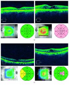
          A multicenter, retrospective study. Data on eyes that underwent anterior or retropupillary iris-claw IOL implantation because of inadequate capsular support secondary to complicated cataract surgery, trauma, and dislocated/opacified IOLs since January 2015 were analyzed. For study inclusion, evaluation results had to be available in the medical records both preoperatively and at 1 and 12 months after implantation. The following parameters were compared between the groups: best-corrected distance visual acuity (BCDVA), spherical and cylindrical refractive error, endothelial cell density (ECD), central macular thickness (CMT), and percentage and type of postoperative complications.

          Results

          In total, 60 eyes of 60 patients aged 73 ± 13 years were included: 28 eyes (47%) involved anterior, and 32 eyes (53%) retropupillary, iris-claw IOL fixations. Preoperatively, the groups were similar in all parameters except for a significantly higher proportion of retropupillary fixations in patients who had previously experienced a closed-globe trauma ( p=0.03). The groups showed comparable improvements in BCDVA after surgery (final BCDVA: 0.34 ± 0.45 vs. 0.37 ± 0.50 logMAR in the anterior and retropupillary placement groups, respectively). During follow-up, no group difference was observed in refractive error or CMT. Both groups experienced similarly marked ECD loss and showed similar incidence of postoperative complications, with cystoid macular edema being the most common complication. Multivariable linear regression showed that BCDVA at 1 month was the best predictor of the final BCDVA.

          Conclusions

          Anterior chamber and posterior chamber iris-claw IOL fixations proved equally effective and safe for aphakic correction in eyes with inadequate capsular support.

          Related collections

          Most cited references18

          • Record: found
          • Abstract: found
          • Article: not found

          Simplified and safe method of sutureless intrascleral posterior chamber intraocular lens fixation: Y-fixation technique.

          We report a new surgical technique that allows intrascleral fixation of a posterior chamber intraocular lens (IOL) without sutures. The Y-fixation technique does not involve complicated intraocular manipulation and achieves safe sutureless fixation. A Y-shaped incision is made in the sclera and a 24-gauge microvitreoretinal (MVR) knife is used to create the sclerotomy instead of a needle. The Y-shaped incision eliminates the need to raise a large lamellar scleral flap and to use fibrin glue because the haptic can be fixed both inside the tunnel and in the groove, and performing the sclerotomy with the 24-gauge MVR knife simplifies extraction of the haptic and improves wound closure. There is no risk of infection from exposure of the haptic on the sclera and no use of fibrin glue. There was significantly less IOL decentration and tilt than with suture fixation.
            Bookmark
            • Record: found
            • Abstract: found
            • Article: not found

            Iris-claw intraocular lenses to correct aphakia in the absence of capsule support.

            To evaluate the indications, postoperative visual efficacy, and complication rate after intraocular implantation of an iris-claw aphakic intraocular lens (IOL). Oxford Eye Hospital, Oxford, United Kingdom. Case series. This chart review comprised eyes with no capsule support that had anterior iris-fixation IOL implantation for aphakia between 2001 and 2009. The study comprised 116 eyes (104 patients). Iris-claw IOLs were inserted during primary lens surgery in 18 eyes (15.5%), during an IOL exchange procedure for dislocated posterior chamber IOLs in 19 eyes (16.4%), and as a secondary procedure in 79 eyes (68.1%). The mean follow-up was 22.4 months (range 3 to 79 months). The final corrected distance visual acuity (CDVA) was 6/12 or better in 68.9% of all eyes and in 47 of 53 eyes (88.7%) with no preoperative comorbidity. Complications included wound leak requiring resuturing in 2.6% of eyes, postoperative intraocular pressure rise in 9.5% of eyes (glaucoma escalation 0.8%), and cystoid macular edema in 7.7% of eyes (0.8% chronic). Iris-claw IOL subluxation occurred in 6.0% of eyes from 5 days to 60 months postoperatively; all the IOLs were repositioned. Corneal decompensation occurred in 1.7% of eyes; 0.8% had retinal detachments. Iris-claw IOL implantation for aphakia gave a good visual outcome and can be used for a wide range of indications. Postoperative complication rates were comparable to, if not better than, those with conventional anterior chamber IOLs. Correct implantation technique is critical in avoiding postoperative IOL subluxation. Copyright © 2011 ASCRS and ESCRS. Published by Elsevier Inc. All rights reserved.
              Bookmark
              • Record: found
              • Abstract: found
              • Article: not found

              Visual outcome and complications after posterior iris-claw aphakic intraocular lens implantation.

              To evaluate the indications, visual outcomes, and complication rate after implantation of a posterior chamber iris-claw aphakic intraocular lens (IOL). Department of Ophthalmology, University Medicine Charité Berlin, Berlin, Germany. Retrospective case series. Eyes without adequate capsule support had posterior chamber iris-claw aphakic IOL implantation (Verisyse/Artisan) between 2005 and 2010. The study comprised 137 eyes (126 patients). The mean follow-up was 5 months (range 1 to 48 months). The IOLs were inserted during primary lens surgery in 10 eyes (7.3%), during an IOL exchange procedure for dislocated posterior chamber IOLs in 95 eyes (69.4%), and as a secondary procedure in 32 aphakic eyes (23.3%). The final mean corrected distance visual acuity (CDVA) (0.38 ± 0.31 [SD] logMAR) was significantly better than preoperatively (0.65 ± 0.58 logMAR) (P < .05). In 128 eyes (93.4%), postoperative refractive errors were within ±2.00 diopters (D) of emmetropia. Complications included slight temporary pupil ovalization in 34 eyes (24.8%), cystoid macular edema in 12 eyes (8.7%), hyphema in 3 eyes (2.1%), early postoperative hypotony in 7 eyes (5.1%) and elevated intraocular pressure in 6 eyes (4.3%), chronic uveitis in 1 eye (0.7%), toxic anterior segment syndrome in 1 eye (0.7%), and endophthalmitis in 1 eye (0.7%). Iris-claw IOL disenclavation occurred in 12 eyes (8.7%); all IOLs could be easily repositioned. The retropupillary iris-claw IOL provided good visual outcomes with a favorable complication rate and can be used for a wide range of indications in eyes without adequate capsule support. No author has a financial or proprietary interest in any material or method mentioned. Copyright © 2012 ASCRS and ESCRS. Published by Elsevier Inc. All rights reserved.
                Bookmark

                Author and article information

                Contributors
                Journal
                J Ophthalmol
                J Ophthalmol
                JOPH
                Journal of Ophthalmology
                Hindawi
                2090-004X
                2090-0058
                2018
                4 November 2018
                : 2018
                : 8463569
                Affiliations
                1Ophthalmology Unit, University Hospital of Parma, Parma, Italy
                2Independent Researcher, Parma, Italy
                3Aditya Jyot Eye Hospital, Wadala, Mumbai, India
                4Department of Ophthalmology, University of Padova, Padova, Italy
                5Eye Clinic, Department of Neurosciences, Biomedicine and Movement Sciences, University of Verona, AOUI-Policlinico G. B. Rossi, Verona, Italy
                Author notes

                Academic Editor: Tamer A. Macky

                Author information
                http://orcid.org/0000-0001-7133-8250
                http://orcid.org/0000-0002-6792-0149
                http://orcid.org/0000-0002-9535-1131
                Article
                10.1155/2018/8463569
                6247566
                30524757
                4de6e4c0-2282-4296-8943-1d7193c0818b
                Copyright © 2018 Paolo Mora et al.

                This is an open access article distributed under the Creative Commons Attribution License, which permits unrestricted use, distribution, and reproduction in any medium, provided the original work is properly cited.

                History
                : 3 July 2018
                : 26 September 2018
                : 8 October 2018
                Categories
                Clinical Study

                Ophthalmology & Optometry
                Ophthalmology & Optometry

                Comments

                Comment on this article