2
views
0
recommends
+1 Recommend
0 collections
    0
    shares
      • Record: found
      • Abstract: found
      • Article: found
      Is Open Access

      Brain functional and effective connectivity based on electroencephalography recordings: A review

      review-article

      Read this article at

      Bookmark
          There is no author summary for this article yet. Authors can add summaries to their articles on ScienceOpen to make them more accessible to a non-specialist audience.

          Abstract

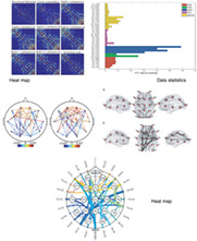
          Functional connectivity and effective connectivity of the human brain, representing statistical dependence and directed information flow between cortical regions, significantly contribute to the study of the intrinsic brain network and its functional mechanism. Many recent studies on electroencephalography (EEG) have been focusing on modeling and estimating brain connectivity due to increasing evidence that it can help better understand various brain neurological conditions. However, there is a lack of a comprehensive updated review on studies of EEG‐based brain connectivity, particularly on visualization options and associated machine learning applications, aiming to translate those techniques into useful clinical tools. This article reviews EEG‐based functional and effective connectivity studies undertaken over the last few years, in terms of estimation, visualization, and applications associated with machine learning classifiers. Methods are explored and discussed from various dimensions, such as either linear or nonlinear, parametric or nonparametric, time‐based, and frequency‐based or time‐frequency‐based. Then it is followed by a novel review of brain connectivity visualization methods, grouped by Heat Map, data statistics, and Head Map, aiming to explore the variation of connectivity across different brain regions. Finally, the current challenges of related research and a roadmap for future related research are presented.

          Abstract

          This article reviews EEG‐based functional and effective connectivity studies undertaken over the last few years, in terms of estimation, visualization, and applications associated with machine learning classifiers. Methods are explored and discussed from various dimensions, such as either linear or nonlinear, parametric, or nonparametric, time‐based, frequency‐based or time‐frequency‐based. Then it is followed by a novel review of brain connectivity visualization methods, grouped by Heat Map, data statistics and Head Map, aiming to explore the variation of connectivity across different brain regions. Finally, the current challenges of related research and a roadmap for future related research are presented.

          Related collections

          Most cited references153

          • Record: found
          • Abstract: found
          • Article: found
          Is Open Access

          The Human Brainnetome Atlas: A New Brain Atlas Based on Connectional Architecture

          The human brain atlases that allow correlating brain anatomy with psychological and cognitive functions are in transition from ex vivo histology-based printed atlases to digital brain maps providing multimodal in vivo information. Many current human brain atlases cover only specific structures, lack fine-grained parcellations, and fail to provide functionally important connectivity information. Using noninvasive multimodal neuroimaging techniques, we designed a connectivity-based parcellation framework that identifies the subdivisions of the entire human brain, revealing the in vivo connectivity architecture. The resulting human Brainnetome Atlas, with 210 cortical and 36 subcortical subregions, provides a fine-grained, cross-validated atlas and contains information on both anatomical and functional connections. Additionally, we further mapped the delineated structures to mental processes by reference to the BrainMap database. It thus provides an objective and stable starting point from which to explore the complex relationships between structure, connectivity, and function, and eventually improves understanding of how the human brain works. The human Brainnetome Atlas will be made freely available for download at http://atlas.brainnetome.org, so that whole brain parcellations, connections, and functional data will be readily available for researchers to use in their investigations into healthy and pathological states.
            Bookmark
            • Record: found
            • Abstract: found
            • Article: not found

            Functional connectome fingerprinting: Identifying individuals based on patterns of brain connectivity

            While fMRI studies typically collapse data from many subjects, brain functional organization varies between individuals. Here, we establish that this individual variability is both robust and reliable, using data from the Human Connectome Project to demonstrate that functional connectivity profiles act as a “fingerprint” that can accurately identify subjects from a large group. Identification was successful across scan sessions and even between task and rest conditions, indicating that an individual’s connectivity profile is intrinsic, and can be used to distinguish that individual regardless of how the brain is engaged during imaging. Characteristic connectivity patterns were distributed throughout the brain, but notably, the frontoparietal network emerged as most distinctive. Furthermore, we show that connectivity profiles predict levels of fluid intelligence; the same networks that were most discriminating of individuals were also most predictive of cognitive behavior. Results indicate the potential to draw inferences about single subjects based on functional connectivity fMRI.
              Bookmark
              • Record: found
              • Abstract: found
              • Article: not found

              Phase lag index: assessment of functional connectivity from multi channel EEG and MEG with diminished bias from common sources.

              To address the problem of volume conduction and active reference electrodes in the assessment of functional connectivity, we propose a novel measure to quantify phase synchronization, the phase lag index (PLI), and compare its performance to the well-known phase coherence (PC), and to the imaginary component of coherency (IC). The PLI is a measure of the asymmetry of the distribution of phase differences between two signals. The performance of PLI, PC, and IC was examined in (i) a model of 64 globally coupled oscillators, (ii) an EEG with an absence seizure, (iii) an EEG data set of 15 Alzheimer patients and 13 control subjects, and (iv) two MEG data sets. PLI and PC were more sensitive than IC to increasing levels of true synchronization in the model. PC and IC were influenced stronger than PLI by spurious correlations because of common sources. All measures detected changes in synchronization during the absence seizure. In contrast to PC, PLI and IC were barely changed by the choice of different montages. PLI and IC were superior to PC in detecting changes in beta band connectivity in AD patients. Finally, PLI and IC revealed a different spatial pattern of functional connectivity in MEG data than PC. The PLI performed at least as well as the PC in detecting true changes in synchronization in model and real data but, at the same token and like-wise the IC, it was much less affected by the influence of common sources and active reference electrodes. Copyright 2007 Wiley-Liss, Inc.
                Bookmark

                Author and article information

                Contributors
                yifan.zhao@cranfield.ac.uk
                Journal
                Hum Brain Mapp
                Hum Brain Mapp
                10.1002/(ISSN)1097-0193
                HBM
                Human Brain Mapping
                John Wiley & Sons, Inc. (Hoboken, USA )
                1065-9471
                1097-0193
                20 October 2021
                1 February 2022
                : 43
                : 2 ( doiID: 10.1002/hbm.v43.2 )
                : 860-879
                Affiliations
                [ 1 ] School of Aerospace, Transport and Manufacturing Cranfield University Cranfield
                [ 2 ] Institute of Geology and Geophysics, Chinese Academy of Sciences Beijing China
                [ 3 ] Department of Automatic Control and Systems Engineering University of Sheffield Sheffield UK
                [ 4 ] School of Automation Science and Electrical Engineering Beihang University Beijing China
                [ 5 ] Department of Neurosurgery Shengjing Hospital of China Medical University Shenyang China
                [ 6 ] Royal Devon and Exeter NHS Foundation Trust Exeter UK
                Author notes
                [*] [* ] Correspondence

                Yifan Zhao, School of Aerospace, Transport and Manufacturing, Cranfield University, Cranfield MK43 0AL, UK.

                Email: yifan.zhao@ 123456cranfield.ac.uk

                Author information
                https://orcid.org/0000-0003-2121-7631
                https://orcid.org/0000-0003-2383-5724
                https://orcid.org/0000-0003-0817-0357
                https://orcid.org/0000-0002-4704-7346
                https://orcid.org/0000-0002-8588-5172
                https://orcid.org/0000-0002-6817-8644
                https://orcid.org/0000-0002-8046-9911
                https://orcid.org/0000-0002-8380-8755
                Article
                HBM25683
                10.1002/hbm.25683
                8720201
                34668603
                4da7f20f-1632-4346-aa36-4f5777d84764
                © 2021 The Authors. Human Brain Mapping published by Wiley Periodicals LLC.

                This is an open access article under the terms of the http://creativecommons.org/licenses/by-nc-nd/4.0/ License, which permits use and distribution in any medium, provided the original work is properly cited, the use is non‐commercial and no modifications or adaptations are made.

                History
                : 10 September 2021
                : 11 May 2021
                : 27 September 2021
                Page count
                Figures: 3, Tables: 3, Pages: 20, Words: 16631
                Funding
                Funded by: National Natural Science Foundation of China , doi 10.13039/501100001809;
                Award ID: 61876015
                Funded by: Beijing Natural Science Foundation, China
                Award ID: 4202040
                Categories
                Review Article
                Review Article
                Custom metadata
                2.0
                February 1, 2022
                Converter:WILEY_ML3GV2_TO_JATSPMC version:6.7.0 mode:remove_FC converted:01.01.2022

                Neurology
                artificial intelligence,brain association,electroencephalogram,machine learning,survey
                Neurology
                artificial intelligence, brain association, electroencephalogram, machine learning, survey

                Comments

                Comment on this article