12
views
0
recommends
+1 Recommend
0 collections
    0
    shares
      • Record: found
      • Abstract: found
      • Article: found
      Is Open Access

      S1PR1 regulates the switch of two angiogenic modes by VE-cadherin phosphorylation in breast cancer

      research-article

      Read this article at

      Bookmark
          There is no author summary for this article yet. Authors can add summaries to their articles on ScienceOpen to make them more accessible to a non-specialist audience.

          Abstract

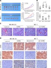
          Angiogenesis in solid tumors is divided into two modes: endothelium-dependent vessel (EDV) and vasculogenic mimicry (VM). Sphingosine-1-phosphate receptor 1 (S1PR1) plays a vital role on EDV in a variety of human tumors. However, the relationship between S1PR1 and VM is not clear. The aim of this study is to investigate S1PR1 on the regulation of EDV and mimicry formation in breast cancer. Here we show that S1PR1 phosphorylates the complex of VE-cadherin to regulate the switch of EDV and mimicry formation. Suppression of S1PR1 impairs EDV, but contributes to the generation of VM, invasion, and metastasis in vivo and vitro. By inhibiting RhoA activation, the S1PR1/VE-cadherin signaling is blocked. S1PR1 controls VE-cadherin expression and EDV via RhoA activation. Moreover, the low expression of S1PR1 correlates with VM and poor prognosis in breast cancer patient. The results show that S1PR1 regulated RhoA activation to accelerate VE-cadherin phosphorylation (Y731), leading to increased EDV and reduced VM in breast cancer. S1PR1 may provide a new thinking direction for antiangiogenic therapy for patients with breast cancer.

          Related collections

          Most cited references39

          • Record: found
          • Abstract: found
          • Article: not found

          Emerging biology of sphingosine-1-phosphate: its role in pathogenesis and therapy.

          Membrane sphingolipids are metabolized to sphingosine-1-phosphate (S1P), a bioactive lipid mediator that regulates many processes in vertebrate development, physiology, and pathology. Once exported out of cells by cell-specific transporters, chaperone-bound S1P is spatially compartmentalized in the circulatory system. Extracellular S1P interacts with five GPCRs that are widely expressed and transduce intracellular signals to regulate cellular behavior, such as migration, adhesion, survival, and proliferation. While many organ systems are affected, S1P signaling is essential for vascular development, neurogenesis, and lymphocyte trafficking. Recently, a pharmacological S1P receptor antagonist has won approval to control autoimmune neuroinflammation in multiple sclerosis. The availability of pharmacological tools as well as mouse genetic models has revealed several physiological actions of S1P and begun to shed light on its pathological roles. The unique mode of signaling of this lysophospholipid mediator is providing novel opportunities for therapeutic intervention, with possibilities to target not only GPCRs but also transporters, metabolic enzymes, and chaperones.
            Bookmark
            • Record: found
            • Abstract: found
            • Article: not found

            Edg-1, the G protein-coupled receptor for sphingosine-1-phosphate, is essential for vascular maturation.

            Sphingolipid signaling pathways have been implicated in many critical cellular events. Sphingosine-1-phosphate (SPP), a sphingolipid metabolite found in high concentrations in platelets and blood, stimulates members of the endothelial differentiation gene (Edg) family of G protein-coupled receptors and triggers diverse effects, including cell growth, survival, migration, and morphogenesis. To determine the in vivo functions of the SPP/Edg signaling pathway, we disrupted the Edg1 gene in mice. Edg1(-/-) mice exhibited embryonic hemorrhage leading to intrauterine death between E12.5 and E14.5. Vasculogenesis and angiogenesis appeared normal in the mutant embryos. However, vascular maturation was incomplete due to a deficiency of vascular smooth muscle cells/pericytes. We also show that Edg-1 mediates an SPP-induced migration response that is defective in mutant cells due to an inability to activate the small GTPase, Rac. Our data reveal Edg-1 to be the first G protein-coupled receptor required for blood vessel formation and show that sphingolipid signaling is essential during mammalian development.
              Bookmark
              • Record: found
              • Abstract: found
              • Article: not found

              Sphingosine-1-phosphate as a ligand for the G protein-coupled receptor EDG-1.

              The sphingolipid metabolite sphingosine-1-phosphate (SPP) has been implicated as a second messenger in cell proliferation and survival. However, many of its biological effects are due to binding to unidentified receptors on the cell surface. SPP activated the heterotrimeric guanine nucleotide binding protein (G protein)-coupled orphan receptor EDG-1, originally cloned as Endothelial Differentiation Gene-1. EDG-1 bound SPP with high affinity (dissociation constant = 8.1 nM) and high specificity. Overexpression of EDG-1 induced exaggerated cell-cell aggregation, enhanced expression of cadherins, and formation of well-developed adherens junctions in a manner dependent on SPP and the small guanine nucleotide binding protein Rho.
                Bookmark

                Author and article information

                Contributors
                +86-13602042200 , xiulanzhao@aliyun.com
                +86-13602111192 , baocunsun@aliyun.com
                Journal
                Cell Death Dis
                Cell Death Dis
                Cell Death & Disease
                Nature Publishing Group UK (London )
                2041-4889
                27 February 2019
                27 February 2019
                March 2019
                : 10
                : 3
                : 200
                Affiliations
                [1 ]ISNI 0000 0000 9792 1228, GRID grid.265021.2, Department of Pathology, , Tianjin Medical University, ; Tianjin, China
                [2 ]ISNI 0000 0004 1757 9434, GRID grid.412645.0, Department of Pathology, , General Hospital of Tianjin Medical University, ; Tianjin, China
                [3 ]ISNI 0000 0004 1798 6427, GRID grid.411918.4, Department of Pathology, , Cancer Hospital of Tianjin Medical University, ; Tianjin, China
                Article
                1411
                10.1038/s41419-019-1411-x
                6393557
                30814488
                4cd55b3f-59a8-489d-8fbd-3f7290a707f6
                © The Author(s) 2019

                Open Access This article is licensed under a Creative Commons Attribution 4.0 International License, which permits use, sharing, adaptation, distribution and reproduction in any medium or format, as long as you give appropriate credit to the original author(s) and the source, provide a link to the Creative Commons license, and indicate if changes were made. The images or other third party material in this article are included in the article’s Creative Commons license, unless indicated otherwise in a credit line to the material. If material is not included in the article’s Creative Commons license and your intended use is not permitted by statutory regulation or exceeds the permitted use, you will need to obtain permission directly from the copyright holder. To view a copy of this license, visit http://creativecommons.org/licenses/by/4.0/.

                History
                : 9 November 2018
                : 27 January 2019
                : 28 January 2019
                Funding
                Funded by: National Natural Science Foundation of China (No. 81572872)
                Funded by: National Natural Science Foundation of China (No.81773076)
                Categories
                Article
                Custom metadata
                © The Author(s) 2019

                Cell biology
                Cell biology

                Comments

                Comment on this article