1
views
0
recommends
+1 Recommend
0 collections
    0
    shares
      • Record: found
      • Abstract: found
      • Article: found
      Is Open Access

      Rediscovering the Rete Ovarii: a secreting auxiliary structure to the ovary

      Preprint
      research-article

      Read this article at

      Bookmark
          There is no author summary for this article yet. Authors can add summaries to their articles on ScienceOpen to make them more accessible to a non-specialist audience.

          Abstract

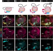
          The rete ovarii (RO) is an appendage of the ovary that has been given little attention. Although the RO appears in drawings of the ovary in early versions of Gray’s Anatomy, it disappeared from recent textbooks, and is often dismissed as a functionless vestige in the adult ovary. Using PAX8 immunostaining and confocal microscopy, we characterized the fetal development of the RO in the context of the ovary. The RO consists of three distinct regions that persist in adult life, the intraovarian rete (IOR), the extraovarian rete (EOR), and the connecting rete (CR). While the cells of the IOR appear to form solid cords within the ovary, the EOR rapidly develops into a convoluted tubular epithelium ending in a distal dilated tip. Cells of the EOR are ciliated and exhibit cellular trafficking capabilities. The CR, connecting the EOR to the IOR, gradually acquires tubular epithelial characteristics by birth. Using microinjections into the distal dilated tip of the EOR, we found that luminal contents flow towards the ovary. Mass spectrometry revealed that the EOR lumen contains secreted proteins potentially important for ovarian function. We show that the cells of the EOR are closely associated with vasculature and macrophages, and are contacted by neuronal projections. The direct proximity of the RO to the ovary and its integration with the extraovarian landscape suggest that it plays an important role in ovary development and homeostasis.

          Related collections

          Most cited references42

          • Record: found
          • Abstract: found
          • Article: not found

          A global double-fluorescent Cre reporter mouse.

          The Cre/loxP system has been used extensively for conditional mutagenesis in mice. Reporters of Cre activity are important for defining the spatial and temporal extent of Cre-mediated recombination. Here we describe mT/mG, a double-fluorescent Cre reporter mouse that expresses membrane-targeted tandem dimer Tomato (mT) prior to Cre-mediated excision and membrane-targeted green fluorescent protein (mG) after excision. We show that reporter expression is nearly ubiquitous, allowing visualization of fluorescent markers in live and fixed samples of all tissues examined. We further demonstrate that mG labeling is Cre-dependent, complementary to mT at single cell resolution, and distinguishable by fluorescence-activated cell sorting. Both membrane-targeted markers outline cell morphology, highlight membrane structures, and permit visualization of fine cellular processes. In addition to serving as a global Cre reporter, the mT/mG mouse may also be used as a tool for lineage tracing, transplantation studies, and analysis of cell morphology in vivo.
            Bookmark
            • Record: found
            • Abstract: found
            • Article: not found

            Neuroepithelial circuit formed by innervation of sensory enteroendocrine cells.

            Satiety and other core physiological functions are modulated by sensory signals arising from the surface of the gut. Luminal nutrients and bacteria stimulate epithelial biosensors called enteroendocrine cells. Despite being electrically excitable, enteroendocrine cells are generally thought to communicate indirectly with nerves through hormone secretion and not through direct cell-nerve contact. However, we recently uncovered in intestinal enteroendocrine cells a cytoplasmic process that we named neuropod. Here, we determined that neuropods provide a direct connection between enteroendocrine cells and neurons innervating the small intestine and colon. Using cell-specific transgenic mice to study neural circuits, we found that enteroendocrine cells have the necessary elements for neurotransmission, including expression of genes that encode pre-, post-, and transsynaptic proteins. This neuroepithelial circuit was reconstituted in vitro by coculturing single enteroendocrine cells with sensory neurons. We used a monosynaptic rabies virus to define the circuit's functional connectivity in vivo and determined that delivery of this neurotropic virus into the colon lumen resulted in the infection of mucosal nerves through enteroendocrine cells. This neuroepithelial circuit can serve as both a sensory conduit for food and gut microbes to interact with the nervous system and a portal for viruses to enter the enteric and central nervous systems.
              Bookmark
              • Record: found
              • Abstract: found
              • Article: not found

              Defining the epithelial stem cell niche in skin.

              Many adult regenerative cells divide infrequently but have high proliferative capacity. We developed a strategy to fluorescently label slow-cycling cells in a cell type-specific fashion. We used this method to purify the label-retaining cells (LRCs) that mark the skin stem cell (SC) niche. We found that these cells rarely divide within their niche but change properties abruptly when stimulated to exit. We determined their transcriptional profile, which, when compared to progeny and other SCs, defines the niche. Many of the >100 messenger RNAs preferentially expressed in the niche encode surface receptors and secreted proteins, enabling LRCs to signal and respond to their environment.
                Bookmark

                Author and article information

                Journal
                bioRxiv
                BIORXIV
                bioRxiv
                Cold Spring Harbor Laboratory
                11 November 2023
                : 2023.11.08.566085
                Affiliations
                [1 ]Department of Cell Biology, Duke University Medical Center, Durham NC 27710
                [2 ]Section of Developmental Biology, Department of Pediatrics, University of Colorado Anschutz Medical Campus, Aurora CO 80045
                Author notes
                [* ]Corresponding author: blanche.capel@ 123456duke.edu

                Author contributions

                DNA, JM, DSL carried out the experiments; DNA carried out the data curating, validating, and formal analysis; DNA, JM, DSL, and BC designed the experiments; DNA, JM, DSL, MB, and BC drafted, revised, and edited the manuscript; JM, MB, and BC acquired funding for the project.

                Author information
                http://orcid.org/0000-0003-0435-4749
                http://orcid.org/0000-0002-2640-1502
                http://orcid.org/0000-0003-0240-5178
                http://orcid.org/0000-0002-3829-0168
                http://orcid.org/0000-0002-6587-0969
                Article
                10.1101/2023.11.08.566085
                10659334
                37986754
                4cce659e-ad45-49b9-b8dc-a16162ec3404

                This work is licensed under a Creative Commons Attribution 4.0 International License, which allows reusers to distribute, remix, adapt, and build upon the material in any medium or format, so long as attribution is given to the creator. The license allows for commercial use.

                History
                Funding
                Funded by: National Institutes of Health
                Award ID: #1R01HD090050-0, #1R01DK1321120, #K99HD103778, #R00HD103778
                Funded by: Duke School of Medicine
                Award ID: #5R01HD039963
                Categories
                Article

                rete ovarii,ovary,proteomics,snare complex,fluid secretion
                rete ovarii, ovary, proteomics, snare complex, fluid secretion

                Comments

                Comment on this article