12
views
0
recommends
+1 Recommend
0 collections
    0
    shares
      • Record: found
      • Abstract: found
      • Article: found
      Is Open Access

      Implementation of a dual-phase grating interferometer for multi-scale characterization of building materials by tunable dark-field imaging

      research-article

      Read this article at

      Bookmark
          There is no author summary for this article yet. Authors can add summaries to their articles on ScienceOpen to make them more accessible to a non-specialist audience.

          Abstract

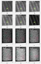
          The multi-scale characterization of building materials is necessary to understand complex mechanical processes, with the goal of developing new more sustainable materials. To that end, imaging methods are often used in materials science to characterize the microscale. However, these methods compromise the volume of interest to achieve a higher resolution. Dark-field (DF) contrast imaging is being investigated to characterize building materials in length scales smaller than the resolution of the imaging system, allowing a direct comparison of features in the nano-scale range and overcoming the scale limitations of the established characterization methods. This work extends the implementation of a dual-phase X-ray grating interferometer (DP-XGI) for DF imaging in a lab-based setup. The interferometer was developed to operate at two different design energies of 22.0 keV and 40.8 keV and was designed to characterize nanoscale-size features in millimeter-sized material samples. The good performance of the interferometer in the low energy range (LER) is demonstrated by the DF retrieval of natural wood samples. In addition, a high energy range (HER) configuration is proposed, resulting in higher mean visibility and good sensitivity over a wider range of correlation lengths in the nanoscale range. Its potential for the characterization of mineral building materials is illustrated by the DF imaging of a Ketton limestone. Additionally, the capability of the DP-XGI to differentiate features in the nanoscale range is proven with the dark-field of Silica nanoparticles at different correlation lengths of calibrated sizes of 106 nm, 261 nm, and 507 nm.

          Related collections

          Most cited references48

          • Record: found
          • Abstract: not found
          • Article: not found

          Phase retrieval and differential phase-contrast imaging with low-brilliance X-ray sources

            Bookmark
            • Record: found
            • Abstract: found
            • Article: not found

            Hard-X-ray dark-field imaging using a grating interferometer.

            Imaging with visible light today uses numerous contrast mechanisms, including bright- and dark-field contrast, phase-contrast schemes and confocal and fluorescence-based methods. X-ray imaging, on the other hand, has only recently seen the development of an analogous variety of contrast modalities. Although X-ray phase-contrast imaging could successfully be implemented at a relatively early stage with several techniques, dark-field imaging, or more generally scattering-based imaging, with hard X-rays and good signal-to-noise ratio, in practice still remains a challenging task even at highly brilliant synchrotron sources. In this letter, we report a new approach on the basis of a grating interferometer that can efficiently yield dark-field scatter images of high quality, even with conventional X-ray tube sources. Because the image contrast is formed through the mechanism of small-angle scattering, it provides complementary and otherwise inaccessible structural information about the specimen at the micrometre and submicrometre length scale. Our approach is fully compatible with conventional transmission radiography and a recently developed hard-X-ray phase-contrast imaging scheme. Applications to X-ray medical imaging, industrial non-destructive testing and security screening are discussed.
              Bookmark
              • Record: found
              • Abstract: found
              • Article: not found

              X-ray phase imaging with a grating interferometer.

              Using a high-efficiency grating interferometer for hard X rays (10-30 keV) and a phase-stepping technique, separate radiographs of the phase and absorption profiles of bulk samples can be obtained from a single set of measurements. Tomographic reconstruction yields quantitative three-dimensional maps of the X-ray refractive index, with a spatial resolution down to a few microns. The method is mechanically robust, requires little spatial coherence and monochromaticity, and can be scaled up to large fields of view, with a detector of correspondingly moderate spatial resolution. These are important prerequisites for use with laboratory X-ray sources.
                Bookmark

                Author and article information

                Contributors
                caori.organista@psi.ch
                Journal
                Sci Rep
                Sci Rep
                Scientific Reports
                Nature Publishing Group UK (London )
                2045-2322
                3 January 2024
                3 January 2024
                2024
                : 14
                : 384
                Affiliations
                [1 ] Radiation Physics Research group, Department Physics and Astronomy, Ghent University, ( https://ror.org/00cv9y106) 9000 Ghent, Belgium
                [2 ]Centre for X-ray Tomography, Ghent University, ( https://ror.org/00cv9y106) 9000 Ghent, Belgium
                [3 ]UGent‑Woodlab, Department of Environment, Faculty of Bioscience Engineering, Ghent University, ( https://ror.org/00cv9y106) 9000 Ghent, Belgium
                [4 ]Pore-Scale Processes in Geomaterials Research Group (PProGRess), Department of Geology, Ghent University, ( https://ror.org/00cv9y106) 9000 Ghent, Belgium
                [5 ]GRID grid.5801.c, ISNI 0000 0001 2156 2780, Institute for Biomedical Engineering, , ETH Zurich, ; 8092 Zurich, Switzerland
                [6 ]GRID grid.5991.4, ISNI 0000 0001 1090 7501, Swiss Light Source, , Paul Scherrer Institute, ; Villigen, 5232 Switzerland
                [7 ] Materials Science and Engineering Division, National Institute of Standards and Technology, ( https://ror.org/05xpvk416) Gaithersburg, MD USA
                Article
                50424
                10.1038/s41598-023-50424-6
                10764912
                38172504
                4ca9520a-e988-4545-81ab-2507ebf88190
                © The Author(s) 2024

                Open Access This article is licensed under a Creative Commons Attribution 4.0 International License, which permits use, sharing, adaptation, distribution and reproduction in any medium or format, as long as you give appropriate credit to the original author(s) and the source, provide a link to the Creative Commons licence, and indicate if changes were made. The images or other third party material in this article are included in the article’s Creative Commons licence, unless indicated otherwise in a credit line to the material. If material is not included in the article’s Creative Commons licence and your intended use is not permitted by statutory regulation or exceeds the permitted use, you will need to obtain permission directly from the copyright holder. To view a copy of this licence, visit http://creativecommons.org/licenses/by/4.0/.

                History
                : 19 September 2023
                : 19 December 2023
                Funding
                Funded by: Fonds Wetenschappelijk Onderzoek (FWO)
                Award ID: 3179I12018
                Award ID: 3179I12018
                Award ID: 3179I12018
                Award Recipient :
                Funded by: FundRef http://dx.doi.org/10.13039/501100001711, Schweizerischer Nationalfonds zur Förderung der Wissenschaftlichen Forschung;
                Award ID: 159263
                Award Recipient :
                Funded by: SwissLOS Lottery Fund of the Kanton of Aargau, Switzerland
                Funded by: Provincie Oost-Vlaanderen (Smart*Light)
                Award ID: 0386
                Award Recipient :
                Categories
                Article
                Custom metadata
                © Springer Nature Limited 2024

                Uncategorized
                engineering,materials science,optics and photonics
                Uncategorized
                engineering, materials science, optics and photonics

                Comments

                Comment on this article

                scite_
                0
                0
                0
                0
                Smart Citations
                0
                0
                0
                0
                Citing PublicationsSupportingMentioningContrasting
                View Citations

                See how this article has been cited at scite.ai

                scite shows how a scientific paper has been cited by providing the context of the citation, a classification describing whether it supports, mentions, or contrasts the cited claim, and a label indicating in which section the citation was made.

                Similar content222

                Most referenced authors324