16
views
0
recommends
+1 Recommend
0 collections
    0
    shares
      • Record: found
      • Abstract: found
      • Article: found
      Is Open Access

      Overdiagnosing of femoroacetabular impingement: correlation between clinical presentation and computed tomography in symptomatic patients Translated title: Sobrediagnóstico do impacto femoroacetabular: correlação entre a clínica e a tomografia computadorizada em pacientes sintomáticos

      research-article

      Read this article at

      Bookmark
          There is no author summary for this article yet. Authors can add summaries to their articles on ScienceOpen to make them more accessible to a non-specialist audience.

          Abstract

          Objective

          To correlate the angles between the acetabulum and the proximal femur in symptomatic patients with femoroacetabular impingement (FAI), using computed tomography (CT).

          Methods

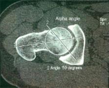
          We retrospectively evaluated 103 hips from 103 patients, using multislice CT to measure the acetabular age, acetabular version (in its supraequatorial portion and in its middle third), femoral neck version, cervical-diaphyseal and alpha angles and the acetabular depth. For the statistical analysis, we used the Pearson correlation coefficient.

          Results

          There were inverse correlations between the following angles: (1) acetabular coverage versus alpha angle ( p = 0.019); (2) acetabular version (supraequatorial) versus alpha angle ( p = 0.049). For patients with femoral anteversion lower than 15 degrees: (1) acetabular version (supraequatorial) versus alpha angle ( p = 0.026); (2) acetabular version (middle third) versus alpha angle ( p = 0.02). For patients with acetabular version (supraequatorial) lower than 10 degrees: (1) acetabular version (supraequatorial) versus alpha angle ( p = 0.004); (2) acetabular version (middle third) versus alpha angle ( p = 0.009).

          Conclusion

          There was a statistically significant inverse correlation between the acetabular version and alpha angles (the smaller the acetabular anteversion angle was, the larger the alpha angle was) in symptomatic patients, thus supporting the hypothesis that FAI occurs when cam and pincer findings due to acetabular retroversion are seen simultaneously, and that the latter alone does not cause FAI, which leads to overdiagnosis in these cases.

          Resumo

          Objetivo

          Correlacionar, por tomografia computadorizada (TC), os ângulos entre o acetábulo e o fêmur proximal em pacientes sintomáticos com impacto femoroacetabular (IFA).

          Métodos

          Avaliamos, retrospectivamente, 103 quadris (103 pacientes) e medimos por TC multislice os ângulos de cobertura acetabular, de versão acetabular (em sua porção supraequatorial e no seu terço médio), de versão do colo femoral, cervicodiafisário, alfa e de profundidade acetabular. Para análise estatística, usamos o coeficiente de correlação de Pearson.

          Resultados

          Houve correlação inversa entre os ângulos: 1) cobertura acetabular versus ângulo alfa (p = 0,019); 2) versão acetabular (supraequatorial) versus ângulo alfa (p = 0,049). Para pacientes com anteversão femoral menor do que 15°: 1) versão acetabular (supraequatorial) versus ângulo alfa (p = 0,026); 2) versão acetabular (terço médio) versus ângulo alfa (p = 0,02). Para pacientes com versão acetabular (supraequatorial) menor do que 10°: 1) versão acetabular (supraequatorial) versus ângulo alfa (p = 0,004); 2) versão acetabular (terço médio) versus ângulo alfa (p = 0,009).

          Conclusão

          Há correlação inversa estatisticamente significativa entre os ângulos de versão acetabular e o ângulo alfa (quanto menor o ângulo de anteversão acetabular, maior o ângulo alfa femoral) em pacientes sintomáticos. Isso reforça a hipótese de que o IFA ocorre quando há simultaneamente os achados de cam e pincer por retroversão acetabular e que esse não causa o IFA isoladamente, o que leva a sobrediagnóstico nesses casos.

          Related collections

          Most cited references60

          • Record: found
          • Abstract: found
          • Article: not found

          Femoroacetabular impingement: a cause for osteoarthritis of the hip.

          A multitude of factors including biochemical, genetic, and acquired abnormalities may contribute to osteoarthritis of the hip. Although the pathomechanism of degenerative process affecting the dysplastic hip is well understood, the exact pathogenesis for idiopathic osteoarthritis has not been established. Based on clinical experience, with more than 600 surgical dislocations of the hip, allowing in situ inspection of the damage pattern and the dynamic proof of its origin, we propose femoroacetabular impingement as a mechanism for the development of early osteoarthritis for most nondysplastic hips. The concept focuses more on motion than on axial loading of the hip. Distinct clinical, radiographic, and intraoperative parameters can be used to confirm the diagnosis of this entity with timely delivery of treatment. Surgical treatment of femoroacetabular impingement focuses on improving the clearance for hip motion and alleviation of femoral abutment against the acetabular rim. It is proposed that early surgical intervention for treatment of femoroacetabular impingement, besides providing relief of symptoms, may decelerate the progression of the degenerative process for this group of young patients.
            Bookmark
            • Record: found
            • Abstract: found
            • Article: not found

            Hip morphology influences the pattern of damage to the acetabular cartilage: femoroacetabular impingement as a cause of early osteoarthritis of the hip.

            Recently, femoroacetabular impingement has been recognised as a cause of early osteoarthritis. There are two mechanisms of impingement: 1) cam impingement caused by a non-spherical head and 2) pincer impingement caused by excessive acetabular cover. We hypothesised that both mechanisms result in different patterns of articular damage. Of 302 analysed hips only 26 had an isolated cam and 16 an isolated pincer impingement. Cam impingement caused damage to the anterosuperior acetabular cartilage with separation between the labrum and cartilage. During flexion, the cartilage was sheared off the bone by the non-spherical femoral head while the labrum remained untouched. In pincer impingement, the cartilage damage was located circumferentially and included only a narrow strip. During movement the labrum is crushed between the acetabular rim and the femoral neck causing degeneration and ossification. Both cam and pincer impingement lead to osteoarthritis of the hip. Labral damage indicates ongoing impingement and rarely occurs alone.
              Bookmark
              • Record: found
              • Abstract: found
              • Article: not found

              The etiology of osteoarthritis of the hip: an integrated mechanical concept.

              The etiology of osteoarthritis of the hip has long been considered secondary (eg, to congenital or developmental deformities) or primary (presuming some underlying abnormality of articular cartilage). Recent information supports a hypothesis that so-called primary osteoarthritis is also secondary to subtle developmental abnormalities and the mechanism in these cases is femoroacetabular impingement rather than excessive contact stress. The most frequent location for femoroacetabular impingement is the anterosuperior rim area and the most critical motion is internal rotation of the hip in 90 degrees flexion. Two types of femoroacetabular impingement have been identified. Cam-type femoroacetabular impingement, more prevalent in young male patients, is caused by an offset pathomorphology between head and neck and produces an outside-in delamination of the acetabulum. Pincer-type femoroacetabular impingement, more prevalent in middle-aged women, is produced by a more linear impact between a local (retroversion of the acetabulum) or general overcoverage (coxa profunda/protrusio) of the acetabulum. The damage pattern is more restricted to the rim and the process of joint degeneration is slower. Most hips, however, show a mixed femoroacetabular impingement pattern with cam predominance. Surgical attempts to restore normal anatomy to avoid femoroacetabular impingement should be performed in the early stage before major cartilage damage is present. Level V, therapeutic study. See the Guidelines for Authors for a complete description of levels of evidence.
                Bookmark

                Author and article information

                Contributors
                Journal
                Rev Bras Ortop
                Rev Bras Ortop
                Revista Brasileira de Ortopedia
                Elsevier
                2255-4971
                16 February 2016
                Mar-Apr 2016
                16 February 2016
                : 51
                : 2
                : 200-207
                Affiliations
                [a ]Imperial Hospital de Caridade, Florianópolis, SC, Brazil
                [b ]Clínica Imagem, Florianópolis, SC, Brazil
                [c ]Hospital Governor Celso Ramos, Florianópolis, SC, Brazil
                [d ]Centro de Pesquisas Oncológicas (CEPON), Florianópolis, SC, Brazil
                Author notes
                [* ] Corresponding author. rpcanella@ 123456md.aaos.org
                Article
                S2255-4971(16)00029-X
                10.1016/j.rboe.2016.02.001
                4812038
                27069890
                4ca66a60-6442-4d3c-b4ff-7d301767ddd3
                © 2015 Sociedade Brasileira de Ortopedia e Traumatologia. Published by Elsevier Editora Ltda. All rights reserved.

                This is an open access article under the CC BY-NC-ND license (http://creativecommons.org/licenses/by-nc-nd/4.0/).

                History
                : 24 March 2015
                : 12 May 2015
                Categories
                Original Article

                hip,femoroacetabular impingement,x-ray computed tomography,quadril,impacto femoroacetabular,tomografia computadorizada por raios x

                Comments

                Comment on this article