2
views
0
recommends
+1 Recommend
0 collections
    0
    shares
      • Record: found
      • Abstract: found
      • Article: found
      Is Open Access

      LGI1-Antibody Associated Autoimmune Encephalitis Complicated by Primary Polydipsia

      letter

      Read this article at

      Bookmark
          There is no author summary for this article yet. Authors can add summaries to their articles on ScienceOpen to make them more accessible to a non-specialist audience.

          Abstract

          Dear Editor Leucine-rich glioma-inactivated 1 (LGI1) antibody encephalitis is one of the subtypes of the autoimmune encephalitis characterized by antibodies against the LGI1 region of the voltage-gated potassium channels.[1] This subtype is characterized by behavioral abnormalities, memory deficits, and classical facio-brachial dystonic seizures besides hyponatremia. We hereby present a typical case of LGI1 associated encephalitis complicated by primary polydipsia previously not reported in the literature. A 28-year-old female was admitted to our department with complaints of memory impairment, irrelevant talking, and abnormal body movements. As reported by her family members, one month back just one week after her engagement, she started questioning about the Mehendi applied on her hands and feet and could not recollect much about her engagement ceremony. Her family members were also concerned about her not recognizing them properly and asking repeatedly for meals even though she would have eaten already. Over the past three weeks she developed abnormal movements of her right arm and face that would last for few seconds and come repeatedly throughout day and night. On examination she was confused, talking irrelevantly and there was no focal neurological deficit. She had multiple episodes of facio-brachial seizures that would last for few seconds only and come repeatedly after a few minutes. Her baseline investigations were normal except for hyponatremia. Because of a subacute encephalopathy, memory deficits, characteristic facio-brachial seizures, and hyponatremia, a provisional diagnosis of LGI1 encephalitis were made. Her spinal fluid analysis revealed 5 cells/μL (all neutrophils) with normal 
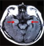
protein (45 mg/dl) and sugar (75 mg/dl with blood sugar of 96 mg/dl). Her MRI brain revealed bilateral symmetrical T2/FLAIR hyperintensities [Figure 1] in mesial temporal lobes and insular cortices with no diffusion restriction or contrast enhancement. Her CSF autoimmune panel was positive for the LGI1 antibody. She was managed with pulse steroid therapy and antiepileptic drugs. However, she did not show any improvement, and instead, she developed polyuria (average 9L/day) and polydipsia (average 8L/day) without polyphagia. Her blood sugar was normal (88 mg/dl) and the main differentials for polyuria and polydipsia were primary polydipsia and diabetes insipidus. However, her serum sodium was persistently low and the water deprivation test did not show a rise in serum osmolality ruling out diabetes insipidus [Table 1]. Hence a diagnosis of LGI1-antibody autoimmune encephalitis complicated by primary polydipsia was made. She was treated by IVIG over 5 days and she showed drastic improvement in encephalopathy, facio-brachial seizures, and osmotic symptoms. She was discharged on steroids and azathioprine and is currently on our follow-up. Figure 1 FLAIR MRI sequence showing hyperintensities in bilateral medial temporal lobes Table 1 Water Deprivation Test Time Urine Osmolality Serum Sodium At 0 hours 130 mOSm/Kg 127 meq/L At 2 hours 277 mOSm/Kg 132 meq/L At 4 hours 397 mOSm/Kg 125 meq/L Limbic encephalitis associated with LGI1 antibodies has certain characteristic features that may make clinical diagnosis easy. Facio-brachial seizures are one such feature; these are dystonic movements of the face and ipsilateral arm that may remain for a few seconds and are usually unilateral but can sometimes involve both sides.[2 3] Prominent psychiatric features like odd behavior, disinhibition, acute psychosis, and memory impairment are other features of this disease.[4] Another characteristic feature of LGI1 associated autoimmune encephalitis is hyponatremia that may be resistant to treatment.[5] Hyponatremia may be seen in 60-80% of patients with LGI1 associated encephalitis and is usually due to SIADH.[6] Imaging may be normal in up to 50% of patients but may reveal T2/FLAIR hyperintensities in medial temporal lobes that are usually unilateral but may also be bilateral.[7] First-line treatment in LGI1 encephalitis is steroids usually in conjunction with intravenous immunoglobulin and/or plasmapheresis. This therapy is usually effective in 80% of patients although the benefit may take time to appear. Our patient had all the characteristic clinical features of LGI1-antibody-associated encephalitis making diagnosis easier further strengthened by a positive CSF LGI1 antibody test. Our patient however did not respond to initial steroid pulse therapy and developed significant polyuria and polydipsia on the 8th day of admission. On the evaluation of these osmotic symptoms, she had persistent hyponatremia with low urine and serum osmolality. The water deprivation test did not increase serum osmolality although there was an increase in urine osmolality thus confirming the diagnosis of primary polydipsia. Primary polydipsia is a condition characterized by excessive consumption of water leading to dilute urine and hyponatremia. Primarily found in psychiatric disease patients like schizophrenics, this disorder can also occur in patients with an organic brain disease like sarcoidosis. This is the first case report of primary polydipsia in a patient with autoimmune encephalitis published in the literature. Our patient was managed with IVIG after which she showed a dramatic response in her seizures and polyuria and polydipsia. As more and more cases of LGI1 encephalitis are reported worldwide newer features of this disease become evident. Primary polydipsia is one such feature that has not been previously reported. Declaration of patient consent The authors certify that they have obtained all appropriate patient consent forms. In the form the patient(s) has/have given his/her/their consent for his/her/their images and other clinical information to be reported in the journal. The patients understand that their names and initials will not be published and due efforts will be made to conceal their identity, but anonymity cannot be guaranteed. Financial support and sponsorship Nil. Conflicts of interest There are no conflicts of interest.

          Related collections

          Most cited references7

          • Record: found
          • Abstract: found
          • Article: not found

          Anti-LGI1 encephalitis: Clinical syndrome and long-term follow-up.

          This nationwide study gives a detailed description of the clinical features and long-term outcome of anti-leucine-rich glioma-inactivated 1 (LGI1) encephalitis.
            Bookmark
            • Record: found
            • Abstract: found
            • Article: found

            Faciobrachial dystonic seizures: the influence of immunotherapy on seizure control and prevention of cognitive impairment in a broadening phenotype.

            Voltage-gated potassium channel complex antibodies, particularly those directed against leucine-rich glioma inactivated 1, are associated with a common form of limbic encephalitis that presents with cognitive impairment and seizures. Faciobrachial dystonic seizures have recently been reported as immunotherapy-responsive, brief, frequent events that often predate the cognitive impairment associated with this limbic encephalitis. However, these observations were made from a retrospective study without serial cognitive assessments. Here, we undertook the first prospective study of faciobrachial dystonic seizures with serial assessments of seizure frequencies, cognition and antibodies in 10 cases identified over 20 months. We hypothesized that (i) faciobrachial dystonic seizures would show a differential response to anti-epileptic drugs and immunotherapy; and that (ii) effective treatment of faciobrachial dystonic seizures would accelerate recovery and prevent the development of cognitive impairment. The 10 cases expand both the known age at onset (28 to 92 years, median 68) and clinical features, with events of longer duration, simultaneously bilateral events, prominent automatisms, sensory aura, and post-ictal fear and speech arrest. Ictal epileptiform electroencephalographic changes were present in three cases. All 10 cases were positive for voltage-gated potassium channel-complex antibodies (346-4515 pM): nine showed specificity for leucine-rich glioma inactivated 1. Seven cases had normal clinical magnetic resonance imaging, and the cerebrospinal fluid examination was unremarkable in all seven tested. Faciobrachial dystonic seizures were controlled more effectively with immunotherapy than anti-epileptic drugs (P = 0.006). Strikingly, in the nine cases who remained anti-epileptic drug refractory for a median of 30 days (range 11-200), the addition of corticosteroids was associated with cessation of faciobrachial dystonic seizures within 1 week in three and within 2 months in six cases. Voltage-gated potassium channel-complex antibodies persisted in the four cases with relapses of faciobrachial dystonic seizures during corticosteroid withdrawal. Time to recovery of baseline function was positively correlated with time to immunotherapy (r = 0.74; P = 0.03) but not time to anti-epileptic drug administration (r = 0.55; P = 0.10). Of 10 cases, the eight cases who received anti-epileptic drugs (n = 3) or no treatment (n = 5) all developed cognitive impairment. By contrast, the two who did not develop cognitive impairment received immunotherapy to treat their faciobrachial dystonic seizures (P = 0.02). In eight cases without clinical magnetic resonance imaging evidence of hippocampal signal change, cross-sectional volumetric magnetic resonance imaging post-recovery, after accounting for age and head size, revealed cases (n = 8) had smaller brain volumes than healthy controls (n = 13) (P < 0.001). In conclusion, faciobrachial dystonic seizures can be prospectively identified as a form of epilepsy with an expanding phenotype. Immunotherapy is associated with excellent control of the frequently anti-epileptic drug refractory seizures, hastens time to recovery, and may prevent the subsequent development of cognitive impairment observed in this study.
              Bookmark
              • Record: found
              • Abstract: found
              • Article: found
              Is Open Access

              Clinical features of limbic encephalitis with LGI1 antibody

              Objective The objective of this study was to analyze the clinical manifestation, course, evolution, image manifestation, and treatments of LGI1 limbic encephalitis (LE). Patients and methods Studies confirmed that LE with the complex antibody of voltage-gated potassium channels is LGI1 LE. Since then, LE cases have been reported. In this study, 10 typical LE cases were searched in PubMed. These cases and one additional case, which we reported herein, were retrospectively analyzed. Results All the patients suffered from recent memory deterioration. The following cases were observed: eight with faciobrachial dystonic seizures (FBDS), six with different kinds of epileptic seizures (four complex partial seizures, one myoclonus seizure, and one generalized tonic–clonic seizure), four with FBDS and different kinds of epileptic seizures at the same time, five with mental disorders (one visual hallucination, one paranoia, one depression, one anxiety, and one dysphoria), five with hyponatremia, and two with sleep disorder. The brain MRI of nine patients revealed abnormalities in the mediotemporal lobe and the hippocampus. The LGI1 antibodies in the blood and/or cerebrospinal fluid (CSF) were positive. The content of the CSF protein of two patients increased slightly. The tumor marker of all the patients was normal, but capitate myxoma was detected in the combined pancreas duct of one patient. Gamma globulin and hormone treatments were administered to nine patients. Of these patients, six received a combination of antiepileptic drugs. The clinical symptoms of all the patients improved. Conclusion LGI1 LE is an autoimmune encephalitis whose clinical manifestations are memory deterioration, FBDS, epileptic seizure, mental disorders, and hyponatremia. Brain MRI shows that this autoimmune disease mainly involves the mediotemporal lobe and the hippocampus. This condition can also be manifested with other autoimmune encephalitis cases but can be rarely associated with tumors. After patients with LGI1 LE receive gamma globulin and hormone treatments, their clinical prognosis is good.
                Bookmark

                Author and article information

                Journal
                Ann Indian Acad Neurol
                Ann Indian Acad Neurol
                AIAN
                Annals of Indian Academy of Neurology
                Wolters Kluwer - Medknow (India )
                0972-2327
                1998-3549
                Jan-Feb 2022
                05 August 2021
                : 25
                : 1
                : 161-163
                Affiliations
                [1]Department of Neurology, Sheri Kashmir Institute of Medical Sciences (SKIMS), Soura, Jammu and Kashmir, India
                Author notes
                Address for correspondence: Dr. Waseem Dar, F 16. New Married Hostel. Block B, SKIMS, Soura, Jammu and Kashmir - 190 011, India. E-mail: drwaseemneurol@ 123456gmail.com
                Article
                AIAN-25-161
                10.4103/aian.AIAN_141_21
                8954302
                4c0365a9-7473-4e8e-8e29-44eeb5dd5575
                Copyright: © 2006 - 2021 Annals of Indian Academy of Neurology

                This is an open access journal, and articles are distributed under the terms of the Creative Commons Attribution-NonCommercial-ShareAlike 4.0 License, which allows others to remix, tweak, and build upon the work non-commercially, as long as appropriate credit is given and the new creations are licensed under the identical terms.

                History
                : 17 February 2021
                : 08 March 2021
                : 18 March 2021
                Categories
                Letters to the Editor

                Neurology
                Neurology

                Comments

                Comment on this article