10
views
0
recommends
+1 Recommend
0 collections
    0
    shares
      • Record: found
      • Abstract: found
      • Article: found
      Is Open Access

      Brachymetacarpia and Brachymetatarsia in Patients with Multiple Hereditary Exostosis

      research-article

      Read this article at

      Bookmark
          There is no author summary for this article yet. Authors can add summaries to their articles on ScienceOpen to make them more accessible to a non-specialist audience.

          Abstract

          Background

          Multiple hereditary exostosis is a common autosomal dominant inherited musculoskeletal disorder that manifests with multiple osteochondromas. The clinical manifestations and pathological characteristics of osteochondromas found in the long bone and genetic alterations related to multiple hereditary exostosis have been widely reported. In this study, we investigated the characteristics of brachymetacarpia and brachymetatarsia associated with multiple hereditary exostosis.

          Methods

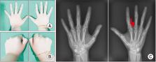
          Of the 133 patients with a diagnosis of multiple hereditary exostosis who were recruited from 2005 to 2018, 101 patients who underwent plain radiography after 10 years of age were included. There were 55 male (54.5%) and 46 female (45.5%) patients. Brachymetacarpia or brachymetatarsia was diagnosed when disruption of the Lièvre parabola connecting the metacarpal or metatarsal heads was observed on plain radiographs. Three orthopedic surgeons individually reviewed hand and foot plain radiographs.

          Results

          Of the 101 patients, 41 patients (40.6%) had more than 1 brachymetacarpia (88 cases) or brachymetatarsia (81 cases). Among 41 cases, 22 (53.7%) were male and 19 (46.3%) were female. The mean age at the time of radiographic evaluation of the hands and feet was 14.6 years (range, 10–63 years). Shortening was most commonly found in the 3rd and 4th metacarpal or metatarsal bones.

          Conclusions

          We found a relatively high incidence of brachymetacarpia and brachymetatarsia in our patients. Physicians should suspect the presence of brachymetacarpia and brachymetatarsia when treating patients with multiple hereditary exostosis.

          Related collections

          Most cited references20

          • Record: found
          • Abstract: found
          • Article: not found

          The natural history of hereditary multiple exostoses.

          We established a database of hereditary multiple exostoses for the state of Washington, on the basis of a retrospective review of the medical records and a clinical evaluation of family members, to determine the prevalence, clinical range of expression, and rate of malignant degeneration. The database comprised forty-six kindreds with 113 affected members; all kindreds had at least one member living in the state of Washington. The over-all prevalence was at least one in 50,000. Approximately 10 per cent of the subjects had no family history of multiple exostoses. With the use of twenty-three pedigrees that demonstrated an adequate multigenerational history for determination of penetrance of the gene, we identified one unaffected individual among twenty-six obligate heterozygotes, a rate of penetrance of 96 per cent. There was no evidence for a substantial reduction of penetrance in female subjects. The median age at the time of the diagnosis in the 113 affected individuals was three years (range, birth to twelve years). In a cohort of eighty-four subjects for whom we had complete information, the clinical range of expression was wide: thirty-three (39 per cent) had an obvious deformity of the forearm, eight (10 per cent) had an inequality in the lengths of the limbs, seven (8 per cent) had an angular deformity of the knee, and two (2 per cent) had a deformity of the ankle. The average number of operations for the patients for whom the operative history was known was two.(ABSTRACT TRUNCATED AT 250 WORDS)
            Bookmark
            • Record: found
            • Abstract: found
            • Article: found
            Is Open Access

            Multiple osteochondromas

            Multiple osteochondromas (MO) is characterised by development of two or more cartilage capped bony outgrowths (osteochondromas) of the long bones. The prevalence is estimated at 1:50,000, and it seems to be higher in males (male-to-female ratio 1.5:1). Osteochondromas develop and increase in size in the first decade of life, ceasing to grow when the growth plates close at puberty. They are pedunculated or sessile (broad base) and can vary widely in size. The number of osteochondromas may vary significantly within and between families, the mean number of locations is 15–18. The majority are asymptomatic and located in bones that develop from cartilage, especially the long bones of the extremities, predominantly around the knee. The facial bones are not affected. Osteochondromas may cause pain, functional problems and deformities, especially of the forearm, that may be reason for surgical removal. The most important complication is malignant transformation of osteochondroma towards secondary peripheral chondrosarcoma, which is estimated to occur in 0.5–5%. MO is an autosomal dominant disorder and is genetically heterogeneous. In almost 90% of MO patients germline mutations in the tumour suppressor genes EXT1 or EXT2 are found. The EXT genes encode glycosyltransferases, catalyzing heparan sulphate polymerization. The diagnosis is based on radiological and clinical documentation, supplemented with, if available, histological evaluation of osteochondromas. If the exact mutation is known antenatal diagnosis is technically possible. MO should be distinguished from metachondromatosis, dysplasia epiphysealis hemimelica and Ollier disease. Osteochondromas are benign lesions and do not affect life expectancy. Management includes removal of osteochondromas when they give complaints. Removed osteochondromas should be examined for malignant transformation towards secondary peripheral chondrosarcoma. Patients should be well instructed and regular follow-up for early detection of malignancy seems justified. For secondary peripheral chondrosarcoma, en-bloc resection of the lesion and its pseudocapsule with tumour-free margins, preferably in a bone tumour referral centre, should be performed.
              Bookmark
              • Record: found
              • Abstract: found
              • Article: not found

              Manifestations and management of osteochondromas: a retrospective analysis of 382 patients.

              Osteochondromas represent the most common primary bone tumours; they reportedly represent 20-50% of all benign bone tumours and 10-15% of all bone tumours. Malignant transformation is their most severe complication. However, deformities and interference with major joint function are the most frequent complaints in patients with hereditary multiple osteochondroma. Treatment should therefore aim not only at surgical resection of the masses but also at prevention of deformities. This article reports observations made on 69 patients with hereditary multiple osteochondroma and 313 patients with solitary osteochondroma, with a mean follow-up of 13.4 years.
                Bookmark

                Author and article information

                Journal
                Clin Orthop Surg
                Clin Orthop Surg
                CIOS
                Clinics in Orthopedic Surgery
                The Korean Orthopaedic Association
                2005-291X
                2005-4408
                December 2020
                18 November 2020
                : 12
                : 4
                : 549-553
                Affiliations
                Department of Orthopaedic Surgery, Chosun University College of Medicine, Gwangju, Korea.
                [* ]Department of Orthopaedic Surgery, Chonnam National University College of Medicine, Gwangju, Korea.
                Author notes
                Correspondence to: Sung Taek Jung, MD. Department of Orthopaedic Surgery, Chonnam National University College of Medicine, 160 Baekseo-ro, Dong-gu, Gwangju 61469, Korea. Tel: +82-62-220-6336, Fax: +82-62-225-7794, stjung@ 123456chonnam.ac.kr
                Article
                10.4055/cios19121
                7683195
                4bede1c3-bd37-4b24-8242-5fff6c328838
                Copyright © 2020 by The Korean Orthopaedic Association

                This is an Open Access article distributed under the terms of the Creative Commons Attribution Non-Commercial License ( http://creativecommons.org/licenses/by-nc/4.0) which permits unrestricted non-commercial use, distribution, and reproduction in any medium, provided the original work is properly cited.

                History
                : 31 July 2019
                : 23 March 2020
                Categories
                Original Article

                Surgery
                multiple hereditary exostosis,deformity,bone development,incidence
                Surgery
                multiple hereditary exostosis, deformity, bone development, incidence

                Comments

                Comment on this article