49
views
0
recommends
+1 Recommend
0 collections
    0
    shares
      • Record: found
      • Abstract: found
      • Article: not found

      Imaging analysis reveals mechanistic differences between first- and second-phase insulin exocytosis

      research-article

      Read this article at

      ScienceOpenPublisherPMC
      Bookmark
          There is no author summary for this article yet. Authors can add summaries to their articles on ScienceOpen to make them more accessible to a non-specialist audience.

          Abstract

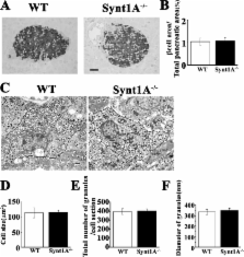
          The mechanism of glucose-induced biphasic insulin release is unknown. We used total internal reflection fluorescence (TIRF) imaging analysis to reveal the process of first- and second-phase insulin exocytosis in pancreatic β cells. This analysis showed that previously docked insulin granules fused at the site of syntaxin (Synt)1A clusters during the first phase; however, the newcomers fused during the second phase external to the Synt1A clusters. To reveal the function of Synt1A in phasic insulin exocytosis, we generated Synt1A-knockout (Synt1A −/−) mice. Synt1A −/− β cells showed fewer previously docked granules with no fusion during the first phase; second-phase fusion from newcomers was preserved. Rescue experiments restoring Synt1A expression demonstrated restoration of granule docking status and fusion events. Inhibition of other syntaxins, Synt3 and Synt4, did not affect second-phase insulin exocytosis. We conclude that the first phase is Synt1A dependent but the second phase is not. This indicates that the two phases of insulin exocytosis differ spatially and mechanistically.

          Related collections

          Most cited references35

          • Record: found
          • Abstract: found
          • Article: not found

          Membrane fusion and exocytosis.

          Membrane fusion involves the merger of two phospholipid bilayers in an aqueous environment. In artificial lipid bilayers, fusion proceeds by means of defined transition states, including hourglass-shaped intermediates in which the proximal leaflets of the fusing membranes are merged whereas the distal leaflets are separate (fusion stalk), followed by the reversible opening of small aqueous fusion pores. Fusion of biological membranes requires the action of specific fusion proteins. Best understood are the viral fusion proteins that are responsible for merging the viral with the host cell membrane during infection. These proteins undergo spontaneous and dramatic conformational changes upon activation. In the case of the paradigmatic fusion proteins of the influenza virus and of the human immunodeficiency virus, an amphiphilic fusion peptide is inserted into the target membrane. The protein then reorients itself, thus forcing the fusing membranes together and inducing lipid mixing. Fusion of intracellular membranes in eukaryotic cells involves several protein families including SNAREs, Rab proteins, and Sec1/Munc-18 related proteins (SM-proteins). SNAREs form a novel superfamily of small and mostly membrane-anchored proteins that share a common motif of about 60 amino acids (SNARE motif). SNAREs reversibly assemble into tightly packed helical bundles, the core complexes. Assembly is thought to pull the fusing membranes closely together, thus inducing fusion. SM-proteins comprise a family of soluble proteins that bind to certain types of SNAREs and prevent the formation of core complexes. Rab proteins are GTPases that undergo highly regulated GTP-GDP cycles. In their GTP form, they interact with specific proteins, the effector proteins. Recent evidence suggests that Rab proteins function in the initial membrane contact connecting the fusing membranes but are not involved in the fusion reaction itself.
            Bookmark
            • Record: found
            • Abstract: found
            • Article: not found

            Hemizygosity at the elastin locus in a developmental disorder, Williams syndrome.

            Williams syndrome (WS) is a developmental disorder affecting connective tissue and the central nervous system. A common feature of WS, supravalvular aortic stenosis, is also a distinct autosomal dominant disorder caused by mutations in the elastin gene. In this study, we identified hemizygosity at the elastin locus using genetic analyses in four familial and five sporadic cases of WS. Fluorescent in situ hybridization and quantitative Southern analyses confirmed these findings, demonstrating inherited and de novo deletions of the elastin gene. These data indicate that deletions involving one elastin allele cause WS and implicate elastin hemizygosity in the pathogenesis of the disease.
              Bookmark
              • Record: found
              • Abstract: found
              • Article: not found

              Membrane fusion.

              Membrane fusion, one of the most fundamental processes in life, occurs when two separate lipid membranes merge into a single continuous bilayer. Fusion reactions share common features, but are catalyzed by diverse proteins. These proteins mediate the initial recognition of the membranes that are destined for fusion and pull the membranes close together to destabilize the lipid/water interface and to initiate mixing of the lipids. A single fusion protein may do everything or assemblies of protein complexes may be required for intracellular fusion reactions to guarantee rigorous regulation in space and time. Cellular fusion machines are adapted to fit the needs of different reactions but operate by similar principles in order to achieve merging of the bilayers.
                Bookmark

                Author and article information

                Journal
                J Cell Biol
                jcb
                The Journal of Cell Biology
                The Rockefeller University Press
                0021-9525
                1540-8140
                21 May 2007
                : 177
                : 4
                : 695-705
                Affiliations
                [1 ]Department of Biochemistry, [2 ]Department of Cell Physiology, [3 ]Department of Anatomy, and [4 ]Department of Clinical Pathology, Kyorin University School of Medicine, Mitaka, Tokyo 181-8611, Japan
                [5 ]Division of Animal Models, Department of Infectious Diseases, Research Institute, International Medical Center of Japan, Tokyo 162-8655, Japan
                [6 ]Department of Medicine, Metabolism and Endocrinology, Juntendo University School of Medicine, Tokyo, 113-8421, Japan
                Author notes

                Correspondence to Shinya Nagamatsu: shinya@ 123456kyorin-u.ac.jp

                Article
                200608132
                10.1083/jcb.200608132
                2064214
                17502420
                4bb09668-9800-4734-a516-f8a0dc2de627
                Copyright © 2007, The Rockefeller University Press
                History
                : 22 August 2006
                : 18 April 2007
                Categories
                Research Articles
                Article

                Cell biology
                Cell biology

                Comments

                Comment on this article