3
views
0
recommends
+1 Recommend
0 collections
    0
    shares
      • Record: found
      • Abstract: found
      • Article: found
      Is Open Access

      Diverse plasma membrane protrusions act as platforms for extracellular vesicle shedding

      review-article
      1 ,
      Journal of Extracellular Vesicles
      John Wiley and Sons Inc.
      actin, extracellular vesicle, filopodium, microvillus, shedding

      Read this article at

      Bookmark
          There is no author summary for this article yet. Authors can add summaries to their articles on ScienceOpen to make them more accessible to a non-specialist audience.

          Abstract

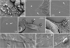
          Plasma membrane curvature is an important factor in the regulation of cellular phenotype and is critical for various cellular activities including the shedding of extracellular vesicles (EV). One of the most striking morphological features of cells is different plasma membrane‐covered extensions supported by actin core such as filopodia and microvilli. Despite the various functions of these extensions are partially unexplained, they are known to facilitate many crucial cellular functions such as migration, adhesion, absorption, and secretion. Due to the rapid increase in the research activity of EVs, there is raising evidence that one of the general features of cellular plasma membrane protrusions is to act as specialized platforms for the budding of EVs. This review will focus on early observations and recent findings supporting this hypothesis, discuss the putative budding and shedding mechanisms of protrusion‐derived EVs and their biological significance.

          Related collections

          Most cited references88

          • Record: found
          • Abstract: found
          • Article: not found

          Shedding light on the cell biology of extracellular vesicles

          Extracellular vesicles are a heterogeneous group of cell-derived membranous structures comprising exosomes and microvesicles, which originate from the endosomal system or which are shed from the plasma membrane, respectively. They are present in biological fluids and are involved in multiple physiological and pathological processes. Extracellular vesicles are now considered as an additional mechanism for intercellular communication, allowing cells to exchange proteins, lipids and genetic material. Knowledge of the cellular processes that govern extracellular vesicle biology is essential to shed light on the physiological and pathological functions of these vesicles as well as on clinical applications involving their use and/or analysis. However, in this expanding field, much remains unknown regarding the origin, biogenesis, secretion, targeting and fate of these vesicles.
            Bookmark
            • Record: found
            • Abstract: found
            • Article: not found

            Exosome-mediated transfer of mRNAs and microRNAs is a novel mechanism of genetic exchange between cells.

            Exosomes are vesicles of endocytic origin released by many cells. These vesicles can mediate communication between cells, facilitating processes such as antigen presentation. Here, we show that exosomes from a mouse and a human mast cell line (MC/9 and HMC-1, respectively), as well as primary bone marrow-derived mouse mast cells, contain RNA. Microarray assessments revealed the presence of mRNA from approximately 1300 genes, many of which are not present in the cytoplasm of the donor cell. In vitro translation proved that the exosome mRNAs were functional. Quality control RNA analysis of total RNA derived from exosomes also revealed presence of small RNAs, including microRNAs. The RNA from mast cell exosomes is transferable to other mouse and human mast cells. After transfer of mouse exosomal RNA to human mast cells, new mouse proteins were found in the recipient cells, indicating that transferred exosomal mRNA can be translated after entering another cell. In summary, we show that exosomes contain both mRNA and microRNA, which can be delivered to another cell, and can be functional in this new location. We propose that this RNA is called "exosomal shuttle RNA" (esRNA).
              Bookmark
              • Record: found
              • Abstract: found
              • Article: found
              Is Open Access

              Extracellular vesicles: Exosomes, microvesicles, and friends

              Cells release into the extracellular environment diverse types of membrane vesicles of endosomal and plasma membrane origin called exosomes and microvesicles, respectively. These extracellular vesicles (EVs) represent an important mode of intercellular communication by serving as vehicles for transfer between cells of membrane and cytosolic proteins, lipids, and RNA. Deficiencies in our knowledge of the molecular mechanisms for EV formation and lack of methods to interfere with the packaging of cargo or with vesicle release, however, still hamper identification of their physiological relevance in vivo. In this review, we focus on the characterization of EVs and on currently proposed mechanisms for their formation, targeting, and function.
                Bookmark

                Author and article information

                Contributors
                kirsi.rilla@uef.fi
                Journal
                J Extracell Vesicles
                J Extracell Vesicles
                10.1002/(ISSN)2001-3078
                JEV2
                Journal of Extracellular Vesicles
                John Wiley and Sons Inc. (Hoboken )
                2001-3078
                17 September 2021
                September 2021
                : 10
                : 11 ( doiID: 10.1002/jev2.v10.11 )
                : e12148
                Affiliations
                [ 1 ] Institute of Biomedicine University of Eastern Finland Kuopio Finland
                Author notes
                [*] [* ] Correspondence

                Kirsi Rilla, Institute of Biomedicine, University of Eastern Finland, Kuopio, Finland.

                Email: kirsi.rilla@ 123456uef.fi

                Author information
                https://orcid.org/0000-0002-7862-5727
                Article
                JEV212148
                10.1002/jev2.12148
                8448080
                34533887
                4b556405-b7c9-46d9-a491-41bd20756e3c
                © 2021 The Authors. Journal of Extracellular Vesicles published by Wiley Periodicals, LLC on behalf of the International Society for Extracellular Vesicles

                This is an open access article under the terms of the http://creativecommons.org/licenses/by/4.0/ License, which permits use, distribution and reproduction in any medium, provided the original work is properly cited.

                History
                : 24 August 2021
                : 02 June 2021
                : 07 September 2021
                Page count
                Figures: 7, Tables: 1, Pages: 13, Words: 8842
                Funding
                Funded by: Academy of Finland GeneCellNano Flagship
                Award ID: 337120
                Funded by: Jane and Aatos Erkko Foundation , doi 10.13039/501100004012;
                Funded by: Mizutani Foundation , doi 10.13039/100009578;
                Categories
                Review Article
                Review Articles
                Custom metadata
                2.0
                September 2021
                Converter:WILEY_ML3GV2_TO_JATSPMC version:6.0.7 mode:remove_FC converted:17.09.2021

                actin,extracellular vesicle,filopodium,microvillus,shedding
                actin, extracellular vesicle, filopodium, microvillus, shedding

                Comments

                Comment on this article