21
views
0
recommends
+1 Recommend
0 collections
    0
    shares
      • Record: found
      • Abstract: found
      • Article: found
      Is Open Access

      Enterohemorrhagic Escherichia coli Reduces Mucus and Intermicrovillar Bridges in Human Stem Cell-Derived Colonoids

      research-article
      1 , 1 , 1 , 2 , 2 , 3 , 4 , 5 , 5 , 1 , 1 ,
      Cellular and Molecular Gastroenterology and Hepatology
      Elsevier
      Human Colonoid Monolayers, Intestinal Organoids, Microvillar Effacement, Serine Protease EspP, A/E, attaching and effacing, BB, brush border, CCS, cold chelating solution, CM, complete medium, 3D, three dimensional, EHEC, enterohemorrhagic Escherichia coli, EM, expansion medium, HCM, human colonoid monolayers, HUS, hemolytic uremic syndrome, IEC, intestinal epithelial cell, LGR5, leucine-rich repeat containing G protein-coupled receptor 5, MLPCDH, mucin-like protocadherin, MUC2, extracellular mucin 2, NHE2, sodium-hydrogen exchanger isoform 2, NHERF3, sodium-hydrogen exchanger regulatory factor 3, PBS, phosphate-buffered saline, PCDH24, protocadherin 24, PCR, polymerase chain reaction, SPATE, serine protease autotransporters of Enterobacteriaceae, Stx, Shiga toxins, TBS, Tris-buffered saline, TER, transepithelial electrical resistance, TJ, tight junction

      Read this article at

      Bookmark
          There is no author summary for this article yet. Authors can add summaries to their articles on ScienceOpen to make them more accessible to a non-specialist audience.

          Abstract

          Background & Aims

          Enterohemorrhagic Escherichia coli (EHEC) causes over 70,000 episodes of foodborne diarrhea annually in the United States. The early sequence of events that precede life-threatening hemorrhagic colitis and hemolytic uremic syndrome is not fully understood due to the initial asymptomatic phase of the disease and the lack of a suitable animal model. We determined the initial molecular events in the interaction between EHEC and human colonic epithelium.

          Methods

          Human colonoids derived from adult proximal colonic stem cells were developed into monolayers to study EHEC-epithelial interactions. Monolayer confluency and differentiation were monitored by transepithelial electrical resistance measurements. The monolayers were apically infected with EHEC, and the progression of epithelial damage over time was assessed using biochemical and imaging approaches.

          Results

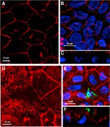
          Human colonoid cultures recapitulate the differential protein expression patterns characteristic of the crypt and surface colonocytes. Mucus-producing differentiated colonoid monolayers are preferentially colonized by EHEC. Upon colonization, EHEC forms characteristic attaching and effacing lesions on the apical surface of colonoid monolayers. Mucin 2, a main component of colonic mucus, and protocadherin 24 (PCDH24), a microvillar resident protein, are targeted by EHEC at early stages of infection. The EHEC-secreted serine protease EspP initiates brush border damage through PCDH24 reduction.

          Conclusions

          Human colonoid monolayers are a relevant pathophysiologic model that allow the study of early molecular events during enteric infections. Colonoid monolayers provide access to both apical and basolateral surfaces, thus providing an advantage over three-dimensional cultures to study host–pathogen interactions in a controllable and tractable manner. EHEC reduces colonic mucus and affects the brush border cytoskeleton in the absence of commensal bacteria.

          Related collections

          Most cited references27

          • Record: found
          • Abstract: found
          • Article: not found

          Development of an enhanced human gastrointestinal epithelial culture system to facilitate patient-based assays.

          The technology for the growth of human intestinal epithelial cells is rapidly progressing. An exciting possibility is that this system could serve as a platform for individualised medicine and research. However, to achieve this goal, human epithelial culture must be enhanced so that biopsies from individuals can be used to reproducibly generate cell lines in a short time frame so that multiple, functional assays can be performed (ie, barrier function and host-microbial interactions).
            Bookmark
            • Record: found
            • Abstract: found
            • Article: not found

            Gene expression patterns of human colon tops and basal crypts and BMP antagonists as intestinal stem cell niche factors.

            Human colonic epithelial cell renewal, proliferation, and differentiation are stringently controlled by numerous regulatory pathways. To identify genetic programs of human colonic epithelial cell differentiation in vivo as well as candidate marker genes that define colonic epithelial stem/progenitor cells and the stem cell niche, we applied gene expression analysis of normal human colon tops and basal crypts by using expression microarrays with 30,000 genes. Nine hundred and sixty-nine cDNA clones were found to be differentially expressed between human colon crypts and tops. Pathway analysis revealed the differential expression of genes involved in cell cycle maintenance and apoptosis, as well as genes in bone morphogenetic protein (BMP), Notch, Wnt, EPH, and MYC signaling pathways. BMP antagonists gremlin 1, gremlin 2, and chordin-like 1 were found to be expressed by colon crypts. In situ hybridization and RT-PCR confirmed that these BMP antagonists are expressed by intestinal cryptal myofibroblasts and smooth muscle cells at the colon crypt. In vitro analysis demonstrated that gremlin 1 partially inhibits Caco-2 cell differentiation upon confluence and activates Wnt signaling in normal rat intestinal epithelial cells. Collectively, the expression data set provides a comprehensive picture of human colonic epithelial cell differentiation. Our study also suggests that BMP antagonists are candidate signaling components that make up the intestinal epithelial stem cell niche.
              Bookmark
              • Record: found
              • Abstract: found
              • Article: not found

              Studies of mucus in mouse stomach, small intestine, and colon. I. Gastrointestinal mucus layers have different properties depending on location as well as over the Peyer's patches.

              Colon has been shown to have a two-layered mucus system where the inner layer is devoid of bacteria. However, a complete overview of the mouse gastrointestinal mucus system is lacking. We now characterize mucus release, thickness, growth over time, adhesive properties, and penetrability to fluorescent beads from stomach to distal colon. Colon displayed spontaneous mucus release and all regions released mucus in response to carbachol and PGE2, except the distal colon and domes of Peyer's patches. Stomach and colon had an inner mucus layer that was adherent to the epithelium. In contrast, the small intestine and Peyer's patches had a single mucus layer that was easily aspirated. The inner mucus layer of the distal colon was not penetrable to beads the size of bacteria and the inner layer of the proximal colon was only partly penetrable. In contrast, the inner mucus layer of stomach was fully penetrable, as was the small intestinal mucus. This suggests a functional organization of the intestinal mucus system, where the small intestine has loose and penetrable mucus that may allow easy penetration of nutrients, in contrast to the stomach, where the mucus provides physical protection, and the colon, where the mucus separates bacteria from the epithelium. This knowledge of the mucus system and its organization improves our understanding of the gastrointestinal tract physiology.
                Bookmark

                Author and article information

                Contributors
                Journal
                Cell Mol Gastroenterol Hepatol
                Cell Mol Gastroenterol Hepatol
                Cellular and Molecular Gastroenterology and Hepatology
                Elsevier
                2352-345X
                22 October 2015
                January 2016
                22 October 2015
                : 2
                : 1
                : 48-62.e3
                Affiliations
                [1 ]Department of Medicine, Division of Gastroenterology and Hepatology, Johns Hopkins University, School of Medicine, Baltimore, Maryland
                [2 ]Department of Microbiology and Immunology, University of Maryland School of Medicine, Baltimore, Maryland
                [3 ]Genetics and Biochemistry Branch, National Institutes of Diabetes and Digestive and Kidney Disease, National Institutes of Health, Bethesda, Maryland
                [4 ]Department of Pathology, Johns Hopkins University School of Medicine, Baltimore, Maryland
                [5 ]Department of Molecular Virology and Microbiology, Baylor College of Medicine, Houston, Texas
                Author notes
                [] Correspondence Address correspondence to: Olga Kovbasnjuk, PhD, Division of Gastroenterology and Hepatology, Johns Hopkins University School of Medicine, 943 Ross Research Building, 720 Rutland Avenue, Baltimore, Maryland 21205.Division of Gastroenterology and HepatologyJohns Hopkins University School of Medicine943 Ross Research Building720 Rutland AvenueBaltimoreMaryland 21205 okovbas1@ 123456jhmi.edu
                Article
                S2352-345X(15)00172-1
                10.1016/j.jcmgh.2015.10.001
                4740923
                26855967
                4b155012-270c-4c18-93eb-f6c847419165
                © 2016 The Authors

                This is an open access article under the CC BY-NC-ND license (http://creativecommons.org/licenses/by-nc-nd/4.0/).

                History
                : 24 August 2015
                : 6 October 2015
                Categories
                Original Research

                human colonoid monolayers,intestinal organoids,microvillar effacement,serine protease espp,a/e, attaching and effacing,bb, brush border,ccs, cold chelating solution,cm, complete medium,3d, three dimensional,ehec, enterohemorrhagic escherichia coli,em, expansion medium,hcm, human colonoid monolayers,hus, hemolytic uremic syndrome,iec, intestinal epithelial cell,lgr5, leucine-rich repeat containing g protein-coupled receptor 5,mlpcdh, mucin-like protocadherin,muc2, extracellular mucin 2,nhe2, sodium-hydrogen exchanger isoform 2,nherf3, sodium-hydrogen exchanger regulatory factor 3,pbs, phosphate-buffered saline,pcdh24, protocadherin 24,pcr, polymerase chain reaction,spate, serine protease autotransporters of enterobacteriaceae,stx, shiga toxins,tbs, tris-buffered saline,ter, transepithelial electrical resistance,tj, tight junction

                Comments

                Comment on this article