31
views
0
recommends
+1 Recommend
0 collections
    0
    shares
      • Record: found
      • Abstract: found
      • Article: not found

      Polo-Like Kinase 1 Directs Assembly of the HsCyk-4 RhoGAP/Ect2 RhoGEF Complex to Initiate Cleavage Furrow Formation

      research-article

      Read this article at

      ScienceOpenPublisherPMC
      Bookmark
          There is no author summary for this article yet. Authors can add summaries to their articles on ScienceOpen to make them more accessible to a non-specialist audience.

          Abstract

          Polo-like kinase 1 promotes assembly of the contractile ring that divides a cell in two by creating a docking site for the RhoA activator Ect2 on the Cyk-4-containing centralspindlin complex at the midzone of the mitotic spindle.

          Abstract

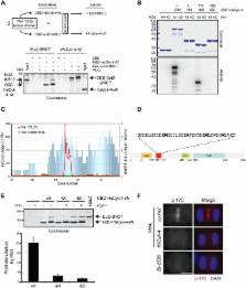
          To complete cell division with high fidelity, cytokinesis must be coordinated with chromosome segregation. Mammalian Polo-like kinase 1, Plk1, may function as a critical link because it is required for chromosome segregation and establishment of the cleavage plane following anaphase onset. A central spindle–localized pool of the RhoGEF Ect2 promotes activation of the small GTPase RhoA, which drives contractile ring assembly at the equatorial cortex. Here, we have investigated how Plk1 promotes the central spindle recruitment of Ect2. Plk1 phosphorylates the noncatalytic N terminus of the RhoGAP HsCyk-4 at the central spindle, creating a phospho-epitope recognized by the BRCA1 C-terminal (BRCT) repeats of Ect2. Failure to phosphorylate HsCyk-4 blocks Ect2 recruitment to the central spindle and the subsequent induction of furrowing. Microtubules, as well as the microtubule-associated protein (MAP) Prc1, facilitate Plk1 phosphorylation of HsCyk-4. Characterization of a phosphomimetic version of HsCyk-4 indicates that Plk1 promotes Ect2 recruitment through multiple targets. Collectively, our data reveal that formation of the HsCyk-4-Ect2 complex is subject to multiple layers of regulation to ensure that RhoA activation occurs between the segregated sister chromatids during anaphase.

          Author Summary

          The plane of cell division in animal cells is determined by the position of the mitotic spindle during early anaphase, but the molecular signaling that leads to proper formation of the division plane is not fully understood. The actin- and myosin-rich contractile ring, which physically divides a cell in two, localizes to the presumptive division plane through the local activation of a molecular switch protein, RhoA. RhoA is activated by Ect2, which binds to the protein complex centralspindlin found on microtubules in the vicinity of the division plane (the midzone microtubules). One critical component of centralspindlin is Cyk-4, a putative negative regulator of RhoA. Here, we have analyzed the mechanisms that are responsible for targeting the RhoA activator Ect2 to the midzone microtubules. We show that Polo-like kinase 1 (Plk1), in part through the microtubule-associated protein Prc1, phosphorylates Cyk-4. Ect2 binds to phosphorylated Cyk-4 and is then able to activate RhoA and induce proper formation of the contractile ring. Our study therefore has elucidated important details of the signaling cascade in animal cells that ensures proper division-plane formation.

          Related collections

          Most cited references43

          • Record: found
          • Abstract: found
          • Article: not found

          BI 2536, a potent and selective inhibitor of polo-like kinase 1, inhibits tumor growth in vivo.

          Fine-mapping of the cell-division cycle, notably the identification of mitotic kinase signaling pathways, provides novel opportunities for cancer-drug discovery. As a key regulator of multiple steps during mitotic progression across eukaryotic species, the serine/threonine-specific Polo-like kinase 1 (Plk1) is highly expressed in malignant cells and serves as a negative prognostic marker in specific human cancer types . Here, we report the discovery of a potent small-molecule inhibitor of mammalian Plk1, BI 2536, which inhibits Plk1 enzyme activity at low nanomolar concentrations. The compound potently causes a mitotic arrest and induces apoptosis in human cancer cell lines of diverse tissue origin and oncogenome signature. BI 2536 inhibits growth of human tumor xenografts in nude mice and induces regression of large tumors with well-tolerated intravenous dose regimens. In treated tumors, cells arrest in prometaphase, accumulate phosphohistone H3, and contain aberrant mitotic spindles. This mitotic arrest is followed by a surge in apoptosis, detectable by immunohistochemistry and noninvasive optical and magnetic resonance imaging. For addressing the therapeutic potential of Plk1 inhibition, BI 2536 has progressed into clinical studies in patients with locally advanced or metastatic cancers.
            Bookmark
            • Record: found
            • Abstract: found
            • Article: not found

            The molecular basis for phosphodependent substrate targeting and regulation of Plks by the Polo-box domain.

            Polo-like kinases (Plks) perform crucial functions in cell-cycle progression and multiple stages of mitosis. Plks are characterized by a C-terminal noncatalytic region containing two tandem Polo boxes, termed the Polo-box domain (PBD), which has recently been implicated in phosphodependent substrate targeting. We show that the PBDs of human, Xenopus, and yeast Plks all recognize similar phosphoserine/threonine-containing motifs. The 1.9 A X-ray structure of a human Plk1 PBD-phosphopeptide complex shows that the Polo boxes each comprise beta6alpha structures that associate to form a 12-stranded beta sandwich domain. The phosphopeptide binds along a conserved, positively charged cleft located at the edge of the Polo-box interface. Mutations that specifically disrupt phosphodependent interactions abolish cell-cycle-dependent localization and provide compelling phenotypic evidence that PBD-phospholigand binding is necessary for proper mitotic progression. In addition, phosphopeptide binding to the PBD stimulates kinase activity in full-length Plk1, suggesting a conformational switching mechanism for Plk regulation and a dual functionality for the PBD.
              Bookmark
              • Record: found
              • Abstract: found
              • Article: not found

              Proteomic screen finds pSer/pThr-binding domain localizing Plk1 to mitotic substrates.

              We have developed a proteomic approach for identifying phosphopeptide binding domains that modulate kinase-dependent signaling pathways. An immobilized library of partially degenerate phosphopeptides biased toward a particular protein kinase phosphorylation motif is used to isolate phospho-binding domains that bind to proteins phosphorylated by that kinase. Applying this approach to cyclin-dependent kinases (Cdks), we identified the polo-box domain (PBD) of the mitotic kinase polo-like kinase 1 (Plk1) as a specific phosphoserine (pSer) or phosphothreonine (pThr) binding domain and determined its optimal binding motif. This motif is present in known Plk1 substrates such as Cdc25, and an optimal phosphopeptide containing the motif disrupted PBD-substrate binding and localization of the PBD to centrosomes. This finding reveals how Plk1 can localize to specific sites within cells in response to Cdk phosphorylation at those sites and provides a structural mechanism for targeting the Plk1 kinase domain to its substrates.
                Bookmark

                Author and article information

                Contributors
                Role: Academic Editor
                Journal
                PLoS Biol
                plos
                plosbiol
                PLoS Biology
                Public Library of Science (San Francisco, USA )
                1544-9173
                1545-7885
                May 2009
                May 2009
                26 May 2009
                : 7
                : 5
                : e1000110
                Affiliations
                [1 ]Department of Molecular Genetics and Cell Biology, The University of Chicago, Chicago, Illinois, United States of America
                [2 ]Cell Division and Aneuploidy Laboratory, Cancer Research UK London Research Institute, Clare Hall Laboratories, Hertfordshire, United Kingdom
                Dana-Farber Cancer Institute, United States of America
                Author notes

                The author(s) have made the following declarations about their contributions: Conceived and designed the experiments: BAW TT MP MG. Performed the experiments: BAW TT MP. Analyzed the data: BAW TT MP MG. Wrote the paper: BAW MP MG.

                Article
                08-PLBI-RA-5050R3
                10.1371/journal.pbio.1000110
                2680334
                19468300
                4ab7cda0-ba0c-496f-9ccc-d8b8f81f41fa
                Wolfe et al. This is an open-access article distributed under the terms of the Creative Commons Attribution License, which permits unrestricted use, distribution, and reproduction in any medium, provided the original author and source are credited.
                History
                : 25 November 2008
                : 31 March 2009
                Page count
                Pages: 15
                Categories
                Research Article
                Cell Biology/Cell Growth and Division
                Cell Biology/Cytoskeleton

                Life sciences
                Life sciences

                Comments

                Comment on this article