0
views
0
recommends
+1 Recommend
0 collections
    0
    shares
      • Record: found
      • Abstract: found
      • Article: found
      Is Open Access

      Expression of guanylate cyclase C in human prefrontal cortex depends on sex and feeding status

      research-article

      Read this article at

      Bookmark
          There is no author summary for this article yet. Authors can add summaries to their articles on ScienceOpen to make them more accessible to a non-specialist audience.

          Abstract

          Introduction

          Guanylate cyclase C (GC-C) has been detected in the rodent brain in neurons of the cerebral cortex, amygdala, midbrain, hypothalamus, and cerebellum.

          Methods

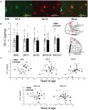
          In this study we determined GC-C protein expression in Brodmann areas (BA) 9, BA10, BA11, and BA32 of the human prefrontal cortex involved in regulation of feeding behavior, as well as in the cerebellar cortex, arcuate nucleus of hypothalamus and substantia nigra in brain samples of human 21 male and 13 female brains by ELISA with postmortem delay < 24 h.

          Results

          GC-C was found in all tested brain areas and it was expressed in neurons of the third cortical layer of BA9. The regulation of GC-C expression by feeding was found in male BA11 and BA10-M, where GC-C expression was in negative correlation to the volume of stomach content during autopsy. In female BA11 there was no correlation detected, while in BA10-M there was even positive correlation. This suggests sex differences in GC-C expression regulation in BA11 and BA10-M. The amount of GC-C was higher in female BA9 only when the death occurred shortly after a meal, while expression of GC-C was higher in BA10-O only when the stomach was empty. The expression of GC-C in female hypothalamus was lower when compared to male hypothalamus only when the stomach was full, suggesting possibly lower satiety effects of GC-C agonists in women.

          Discussion

          These results point toward the possible role of GC-C in regulation of feeding behavior. Since, this is first study of GC-C regulation and its possible function in prefrontal cortex, to determine exact role of GC-C in different region of prefrontal cortex, especially in humans, need further studies.

          Related collections

          Most cited references62

          • Record: found
          • Abstract: found
          • Article: not found

          Temporal dynamics and genetic control of transcription in the human prefrontal cortex.

          Previous investigations have combined transcriptional and genetic analyses in human cell lines, but few have applied these techniques to human neural tissue. To gain a global molecular perspective on the role of the human genome in cortical development, function and ageing, we explore the temporal dynamics and genetic control of transcription in human prefrontal cortex in an extensive series of post-mortem brains from fetal development through ageing. We discover a wave of gene expression changes occurring during fetal development which are reversed in early postnatal life. One half-century later in life, this pattern of reversals is mirrored in ageing and in neurodegeneration. Although we identify thousands of robust associations of individual genetic polymorphisms with gene expression, we also demonstrate that there is no association between the total extent of genetic differences between subjects and the global similarity of their transcriptional profiles. Hence, the human genome produces a consistent molecular architecture in the prefrontal cortex, despite millions of genetic differences across individuals and races. To enable further discovery, this entire data set is freely available (from Gene Expression Omnibus: accession GSE30272; and dbGaP: accession phs000417.v1.p1) and can also be interrogated via a biologist-friendly stand-alone application (http://www.libd.org/braincloud).
            Bookmark
            • Record: found
            • Abstract: found
            • Article: not found

            The organization of networks within the orbital and medial prefrontal cortex of rats, monkeys and humans.

            This paper reviews architectonic subdivisions and connections of the orbital and medial prefrontal cortex (OMPFC) in rats, monkeys and humans. Cortico-cortical connections provide the basis for recognition of 'medial' and 'orbital' networks within the OMPFC. These networks also have distinct connections with structures in other parts of the brain. The orbital network receives sensory inputs from several modalities, including olfaction, taste, visceral afferents, somatic sensation and vision, which appear to be especially related to food or eating. In contrast, the medial network provides the major cortical output to visceromotor structures in the hypothalamus and brainstem. The two networks have distinct connections with areas of the striatum and mediodorsal thalamus. In particular, projections to the nucleus accumbens and the adjacent ventromedial caudate and putamen arise predominantly from the medial network. Both networks also have extensive connections with limbic structures. Based on these and other observations, the OMPFC appears to function as a sensory-visceromotor link, especially for eating. This linkage appears to be critical for the guidance of reward-related behavior and for setting of mood. Imaging and histological observations on human brains indicate that clinical depressive disorders are associated with specific functional and cellular changes in the OMPFC, including activity and volume changes, and specific changes in the number of glial cells.
              Bookmark
              • Record: found
              • Abstract: found
              • Article: not found

              Architectonic subdivision of the human orbital and medial prefrontal cortex.

              The structure of the human orbital and medial prefrontal cortex (OMPFC) was investigated using five histological and immunohistochemical stains and was correlated with a previous analysis in macaque monkeys [Carmichael and Price (1994) J. Comp. Neurol. 346:366-402]. A cortical area was recognized if it was distinct with at least two stains and was found in similar locations in different brains. All of the areas recognized in the macaque OMPFC have counterparts in humans. Areas 11, 13, and 14 were subdivided into areas 11m, 11l, 13a, 13b, 13m, 13l, 14r, and 14c. Within area 10, the region corresponding to area 10m in monkeys was divided into 10m and 10r, and area 10o (orbital) was renamed area 10p (polar). Areas 47/12r, 47/12m, 47/12l, and 47/12s occupy the lateral orbital cortex, corresponding to monkey areas 12r, 12m, 12l, and 12o. The agranular insula (areas Iam, Iapm, Iai, and Ial) extends onto the caudal orbital surface and into the horizontal ramus of the lateral sulcus. The growth of the frontal pole in humans has pushed area 25 and area 32pl, which corresponds to the prelimbic area 32 in Brodmann's monkey brain map, caudal and ventral to the genu of the corpus callosum. Anterior cingulate areas 24a and 24b also extend ventral to the genu of the corpus callosum. Area 32ac, corresponding to the dorsal anterior cingulate area 32 in Brodmann's human brain map, is anterior and dorsal to the genu. The parallel organization of the OMPFC in monkeys and humans allows experimental data from monkeys to be applied to studies of the human cortex. Copyright 2003 Wiley-Liss, Inc.
                Bookmark

                Author and article information

                Contributors
                Role: Role: Role: Role: Role:
                Role: Role: Role: Role:
                Role: Role: Role: Role:
                Role: Role: Role: Role:
                Role: Role: Role: Role: Role:
                Role: Role: Role: Role: Role: Role:
                URI : http://loop.frontiersin.org/people/1351011/overviewRole: Role: Role: Role: Role: Role: Role:
                URI : http://loop.frontiersin.org/people/2613414/overviewRole: Role: Role: Role: Role: Role: Role: Role: Role: Role: Role: Role:
                Journal
                Front Mol Neurosci
                Front Mol Neurosci
                Front. Mol. Neurosci.
                Frontiers in Molecular Neuroscience
                Frontiers Media S.A.
                1662-5099
                22 May 2024
                2024
                : 17
                : 1361089
                Affiliations
                [1] 1Laboratory for Cellular Neurophysiology, Croatian Institute for Brain Research, School of Medicine, University of Zagreb , Zagreb, Croatia
                [2] 2Centre of Excellence for Basic, Clinical and Translational Neuroscience, School of Medicine, University of Zagreb , Zagreb, Croatia
                [3] 3Department of Physiology, School of Medicine, University of Zagreb , Zagreb, Croatia
                [4] 4Institute for Forensic Medicine, School of Medicine, University of Zagreb , Zagreb, Croatia
                [5] 5Department of Anatomy and Clinical Anatomy, School of Medicine, University of Zagreb , Zagreb, Croatia
                Author notes

                Edited by: Marija Heffer, Josip Juraj Strossmayer University of Osijek, Croatia

                Reviewed by: Anna Kozłowska, University of Warmia and Mazury in Olsztyn, Poland

                Branka Hrvoj Mihic, University of California, San Diego, United States

                *Correspondence: Aleksandra Dugandžić aleksandra.dugandzic@ 123456mef.hr
                Article
                10.3389/fnmol.2024.1361089
                11150535
                38840774
                496b653c-b906-432e-982d-ba3cd75c45be
                Copyright © 2024 Ratko, Crljen, Tkalčić, Mažuranić, Bubalo, Škavić, Banovac and Dugandžić.

                This is an open-access article distributed under the terms of the Creative Commons Attribution License (CC BY). The use, distribution or reproduction in other forums is permitted, provided the original author(s) and the copyright owner(s) are credited and that the original publication in this journal is cited, in accordance with accepted academic practice. No use, distribution or reproduction is permitted which does not comply with these terms.

                History
                : 24 December 2023
                : 30 April 2024
                Page count
                Figures: 5, Tables: 2, Equations: 0, References: 62, Pages: 12, Words: 8135
                Funding
                Funded by: Hrvatska Zaklada za Znanost, doi 10.13039/501100004488;
                Award ID: IP-2018-01- 7416
                Funded by: Sveučilište u Zagrebu, doi 10.13039/100008993;
                Award ID: 10106-22-3100
                The author(s) declare financial support was received for the research, authorship, and/or publication of this article. This publication was co-financed by the European Union through the European Regional Development Fund, Operational Programme Competitiveness and Cohesion, Grant Agreement No. KK.01.1.1.01.0007, CoRENeuro, and by the Croatian Science Foundation Research Grant (IP-2018-01- 7416).
                Categories
                Molecular Neuroscience
                Original Research
                Custom metadata
                Neuroplasticity and Development

                Neurosciences
                human prefrontal cortex,cerebellum,hypothalamus,substantia nigra,feeding
                Neurosciences
                human prefrontal cortex, cerebellum, hypothalamus, substantia nigra, feeding

                Comments

                Comment on this article