28
views
0
recommends
+1 Recommend
0 collections
    0
    shares
      • Record: found
      • Abstract: found
      • Article: found
      Is Open Access

      Neurochemical evidence supporting dopamine D1–D2 receptor heteromers in the striatum of the long-tailed macaque: changes following dopaminergic manipulation

      research-article

      Read this article at

      Bookmark
          There is no author summary for this article yet. Authors can add summaries to their articles on ScienceOpen to make them more accessible to a non-specialist audience.

          Abstract

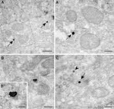
          Although it has long been widely accepted that dopamine receptor types D1 and D2 form GPCR heteromers in the striatum, the presence of D1–D2 receptor heteromers has been recently challenged. In an attempt to properly characterize D1–D2 receptor heteromers, here we have used the in situ proximity ligation assay (PLA) in striatal sections comprising the caudate nucleus, the putamen and the core and shell territories of the nucleus accumbens. Experiments were carried out in control macaques as well as in MPTP-treated animals (with and without dyskinesia). Obtained data support the presence of D1–D2 receptor heteromers within all the striatal subdivisions, with the highest abundance in the accumbens shell. Dopamine depletion by MPTP resulted in an increase of D1–D2 density in caudate and putamen which was normalized by levodopa treatment. Two different sizes of heteromers were consistently found, thus suggesting that besides individual heteromers, D1–D2 receptor heteromers are sometimes organized in macromolecular complexes made of a number of D1–D2 heteromers. Furthermore, the PLA technique was combined with different neuronal markers to properly characterize the identities of striatal neurons expressing D1–D2 heteromers. We have found that striatal projection neurons giving rise to either the direct or the indirect basal ganglia pathways expressed D1–D2 heteromers. Interestingly, macromolecular complexes of D1–D2 heteromers were only found within cholinergic interneurons. In summary, here we provide overwhelming proof that D1 and D2 receptors form heteromeric complexes in the macaque striatum, thus representing a very appealing target for a number of brain diseases involving dopamine dysfunction.

          Related collections

          Most cited references59

          • Record: found
          • Abstract: found
          • Article: not found

          Characterizing proteins and their interactions in cells and tissues using the in situ proximity ligation assay.

          The activity of proteins is typically regulated by secondary modifications and by interactions with other partners, resulting in the formation of protein complexes whose functions depend on the participating proteins. Accordingly, it is of central importance to monitor the presence of interaction complexes as well as their localization, thus providing information about the types of cells where the proteins are located and in what sub-cellular compartment these interactions occur. Several methods for visualizing protein interactions in situ have been developed during the last decade. These methods in most cases involve genetic constructs, and they have been successfully used in assays of living cell maintained in tissue culture, but they cannot easily be implemented in studies of clinical specimens. For such samples, affinity reagents like antibodies can be used to target the interacting proteins. In this review we will describe the in situ proximity ligation assays (in situ PLA), a method that is suitable for visualizing protein interactions in both tissue sections and in vitro cell lines, and we discuss research tasks when this or other method may be selected.
            Bookmark
            • Record: found
            • Abstract: found
            • Article: not found

            Dopamine receptors - IUPHAR Review 13.

            The variety of physiological functions controlled by dopamine in the brain and periphery is mediated by the D1, D2, D3, D4 and D5 dopamine GPCRs. Drugs acting on dopamine receptors are significant tools for the management of several neuropsychiatric disorders including schizophrenia, bipolar disorder, depression and Parkinson's disease. Recent investigations of dopamine receptor signalling have shown that dopamine receptors, apart from their canonical action on cAMP-mediated signalling, can regulate a myriad of cellular responses to fine-tune the expression of dopamine-associated behaviours and functions. Such signalling mechanisms may involve alternate G protein coupling or non-G protein mechanisms involving ion channels, receptor tyrosine kinases or proteins such as β-arrestins that are classically involved in GPCR desensitization. Another level of complexity is the growing appreciation of the physiological roles played by dopamine receptor heteromers. Applications of new in vivo techniques have significantly furthered the understanding of the physiological functions played by dopamine receptors. Here we provide an update of the current knowledge regarding the complex biology, signalling, physiology and pharmacology of dopamine receptors.
              Bookmark
              • Record: found
              • Abstract: found
              • Article: not found

              Opposing patterns of signaling activation in dopamine D1 and D2 receptor-expressing striatal neurons in response to cocaine and haloperidol.

              Psychostimulants and other drugs of abuse activate extracellular signal-regulated kinase (ERK) in the striatum, through combined stimulation of dopamine D(1) receptors (D1Rs) and glutamate NMDA receptors. Antipsychotic drugs activate similar signaling proteins in the striatum by blocking dopamine D(2) receptors (D2Rs). However, the neurons in which these pathways are activated by psychotropic drugs are not precisely identified. We used transgenic mice, in which enhanced green fluorescent protein (EGFP) expression was driven by D1R promoter (drd1a-EGFP) or D2R promoter (drd2-EGFP). We confirmed the expression of drd1a-EGFP in striatonigral and drd2-EGFP in striatopallidal neurons. Drd2-EGFP was also expressed in cholinergic interneurons, whereas no expression of either promoter was detected in GABAergic interneurons. Acute cocaine treatment increased phosphorylation of ERK and its direct or indirect nuclear targets, mitogen- and stress-activated kinase-1 (MSK1) and histone H3, exclusively in D1R-expressing output neurons in the dorsal striatum and nucleus accumbens. Cocaine-induced expression of c-Fos and Zif268 predominated in D1R-expressing neurons but was also observed in D2R-expressing neurons. One week after repeated cocaine administration, cocaine-induced signaling responses were decreased, with the exception of enhanced ERK phosphorylation in dorsal striatum. The responses remained confined to D1R neurons. In contrast, acute haloperidol injection activated phosphorylation of ERK, MSK1, and H3 only in D2R neurons and induced c-fos and zif268 predominantly in these neurons. Our results demonstrate that cocaine and haloperidol specifically activate signaling pathways in two completely segregated populations of striatal output neurons, providing direct evidence for the selective mechanisms by which these drugs exert their long-term effects.
                Bookmark

                Author and article information

                Contributors
                +34 948 194 700 x 2002 , jlanciego@unav.es
                Journal
                Brain Struct Funct
                Brain Struct Funct
                Brain Structure & Function
                Springer Berlin Heidelberg (Berlin/Heidelberg )
                1863-2653
                1863-2661
                9 September 2016
                9 September 2016
                2017
                : 222
                : 4
                : 1767-1784
                Affiliations
                [1 ]ISNI 0000000419370271, GRID grid.5924.a, Department of Neurosciences, , Center for Applied Medical Research (CIMA), University of Navarra, ; Pio XII Avenue 55, 31008 Pamplona, Spain
                [2 ]ISNI 0000 0004 1762 4012, GRID grid.418264.d, , Centro de Investigación Biomédica en Red sobre Enfermedades Neurodegenerativas (CIBERNED), ; Madrid, Spain
                [3 ]ISNI 0000000109410645, GRID grid.11794.3a, Department of Morphological Sciences, , University of Santiago de Compostela, ; Santiago De Compostela, Spain
                [4 ]ISNI 0000 0001 2157 2938, GRID grid.17063.33, Campbell Family Mental Health Research Institute, Centre for Addiction and Mental Health, Departments of Medicine and Pharmacology, , University of Toronto, ; Toronto, ON Canada
                [5 ]ISNI 0000 0004 1937 0247, GRID grid.5841.8, Department of Biochemistry and Molecular Biology, , University of Barcelona, ; Barcelona, Spain
                Article
                1306
                10.1007/s00429-016-1306-x
                5406426
                27612857
                47e8636a-2b7b-42eb-b568-9ac9afb2d993
                © The Author(s) 2016

                Open AccessThis article is distributed under the terms of the Creative Commons Attribution 4.0 International License ( http://creativecommons.org/licenses/by/4.0/), which permits unrestricted use, distribution, and reproduction in any medium, provided you give appropriate credit to the original author(s) and the source, provide a link to the Creative Commons license, and indicate if changes were made.

                History
                : 20 June 2016
                : 6 September 2016
                Funding
                Funded by: FundRef http://dx.doi.org/10.13039/501100003329, Ministerio de Economía y Competitividad;
                Award ID: BFU2012-37907
                Award ID: SAF2012-039875-C02-01
                Award Recipient :
                Funded by: centro de investigacion biomedica en red de enfermedades neurodegenerativas
                Award ID: CB06/05/0006
                Award Recipient :
                Funded by: FundRef http://dx.doi.org/10.13039/100008666, Fundació la Marató de TV3;
                Award ID: 20141331
                Award ID: 20141330
                Award Recipient :
                Funded by: Jon Zarandona
                Categories
                Original Article
                Custom metadata
                © Springer-Verlag Berlin Heidelberg 2017

                Neurology
                basal ganglia,putamen,caudate nucleus,dyskinesia,mptp
                Neurology
                basal ganglia, putamen, caudate nucleus, dyskinesia, mptp

                Comments

                Comment on this article