31
views
0
recommends
+1 Recommend
0 collections
    0
    shares
      • Record: found
      • Abstract: found
      • Article: found
      Is Open Access

      Clinical Potential and Current Progress of Dental Pulp Stem Cells for Various Systemic Diseases in Regenerative Medicine: A Concise Review

      review-article

      Read this article at

      Bookmark
          There is no author summary for this article yet. Authors can add summaries to their articles on ScienceOpen to make them more accessible to a non-specialist audience.

          Abstract

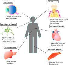
          Dental pulp stem cells (DPSCs) are mesenchymal stem cells (MSCs) that have multipotent differentiation and a self-renewal ability. They have been useful not only for dental diseases, but also for systemic diseases. Extensive studies have suggested that DPSCs are effective for various diseases, such as spinal cord injuries, Parkinson’s disease, Alzheimer’s disease, cerebral ischemia, myocardial infarction, muscular dystrophy, diabetes, liver diseases, eye diseases, immune diseases, and oral diseases. DPSCs have the potential for use in a cell-therapeutic paradigm shift to treat these diseases. It has also been reported that DPSCs have higher regenerative potential than the bone marrow-derived mesenchymal stem cells known as representative MSCs. Therefore, DPSCs have recently gathered much attention. In this review, the therapeutic potential of DPSCs, the latest progress in the pre-clinical study for treatment of these various systemic diseases, and the clinical applications of DPSCs in regenerative medicine, are all summarized. Although challenges, including mechanisms of the effects and establishment of cell processing and transplantation methods for clinical use, still remain, DPSCs could be promising stem cells sources for various clinical applications, because of their easy isolation by a noninvasive procedure without ethical concerns.

          Related collections

          Most cited references75

          • Record: found
          • Abstract: found
          • Article: not found

          Characterization of the apical papilla and its residing stem cells from human immature permanent teeth: a pilot study.

          Mesenchymal stem cells (MSCs) have been isolated from the pulp tissue of permanent teeth (dental pulp stem cells or DPSCs) and deciduous teeth (stem cells from human exfoliated deciduous teeth). We recently discovered another type of MSCs in the apical papilla of human immature permanent teeth termed stem cells from the apical papilla (SCAP). Here, we further characterized the apical papilla tissue and stem cell properties of SCAP using histologic, immunohistochemical, and immunocytofluorescent analyses. We found that the apical papilla is distinctive to the pulp in terms of containing less cellular and vascular components than those in the pulp. Cells in the apical papilla proliferated 2- to 3-fold greater than those in the pulp in organ cultures. Both SCAP and DPSCs were as potent in osteo/dentinogenic differentiation as MSCs from bone marrows, whereas they were weaker in adipogenic potential. The immunophenotype of SCAP is similar to that of DPSCs on the osteo/dentinogenic and growth factor receptor gene profiles. Double-staining experiments showed that STRO-1 coexpressed with dentinogenic markers such as bone sialophosphoprotein, osteocalcin, and growth factors FGFR1 and TGFbetaRI in cultured SCAP. Additionally, SCAP express a wide variety of neurogenic markers such as nestin and neurofilament M upon stimulation with a neurogenic medium. We conclude that SCAP are similar to DPSCs but a distinct source of potent dental stem/progenitor cells. Their implications in root development and apexogenesis are discussed.
            Bookmark
            • Record: found
            • Abstract: found
            • Article: not found

            Fate of the mammalian cranial neural crest during tooth and mandibular morphogenesis.

            Neural crest cells are multipotential stem cells that contribute extensively to vertebrate development and give rise to various cell and tissue types. Determination of the fate of mammalian neural crest has been inhibited by the lack of appropriate markers. Here, we make use of a two-component genetic system for indelibly marking the progeny of the cranial neural crest during tooth and mandible development. In the first mouse line, Cre recombinase is expressed under the control of the Wnt1 promoter as a transgene. Significantly, Wnt1 transgene expression is limited to the migrating neural crest cells that are derived from the dorsal CNS. The second mouse line, the ROSA26 conditional reporter (R26R), serves as a substrate for the Cre-mediated recombination. Using this two-component genetic system, we have systematically followed the migration and differentiation of the cranial neural crest (CNC) cells from E9.5 to 6 weeks after birth. Our results demonstrate, for the first time, that CNC cells contribute to the formation of condensed dental mesenchyme, dental papilla, odontoblasts, dentine matrix, pulp, cementum, periodontal ligaments, chondrocytes in Meckel's cartilage, mandible, the articulating disc of temporomandibular joint and branchial arch nerve ganglia. More importantly, there is a dynamic distribution of CNC- and non-CNC-derived cells during tooth and mandibular morphogenesis. These results are a first step towards a comprehensive understanding of neural crest cell migration and differentiation during mammalian craniofacial development. Furthermore, this transgenic model also provides a new tool for cell lineage analysis and genetic manipulation of neural-crest-derived components in normal and abnormal embryogenesis.
              Bookmark
              • Record: found
              • Abstract: found
              • Article: not found

              Hypoimmunogenic derivatives of induced pluripotent stem cells evade immune rejection in fully immunocompetent allogeneic recipients

              Autologous induced pluripotent stem cells (iPSCs) constitute an unlimited cell source for patient-specific cell-based organ repair strategies. However, their generation and subsequent differentiation into specific cells or tissues entail cell line-specific manufacturing challenges and form a lengthy process that precludes acute treatment modalities. These shortcomings could be overcome by using prefabricated allogeneic cell or tissue products, but the vigorous immune response against histo-incompatible cells has prevented the successful implementation of this approach. Here we show that both mouse and human iPSCs lose their immunogenicity when major histocompatibility complex (MHC) class I and II genes are inactivated and CD47 is over-expressed. These hypoimmunogenic iPSCs retain their pluripotent stem cell potential and differentiation capacity. Endothelial cells, smooth muscle cells, and cardiomyocytes derived from hypoimmunogenic mouse or human iPSCs reliably evade immune rejection in fully MHC-mismatched allogeneic recipients and survive long-term without the use of immunosuppression. These findings suggest that hypoimmunogenic cell grafts can be engineered for universal transplantation.
                Bookmark

                Author and article information

                Journal
                Int J Mol Sci
                Int J Mol Sci
                ijms
                International Journal of Molecular Sciences
                MDPI
                1422-0067
                06 March 2019
                March 2019
                : 20
                : 5
                : 1132
                Affiliations
                [1 ]Department of Oral Implantology, Osaka Dental University, 1-5-17 Otemae Chuo-ku, Osaka 540-0008, Japan; yamada-s@ 123456cc.osaka-dent.ac.jp (S.N.-Y.); kusano-k@ 123456cc.osaka-dent.ac.jp (K.K.); baba-s@ 123456cc.osaka-dent.ac.jp (S.B.)
                [2 ]Department of Infectious Diseases and Applied Immunology, IMSUT Hospital of The Institute of Medical Science, The University of Tokyo, 4-6-1 Shiroganedai, Minato-ku, Tokyo 108-8639, Japan
                Author notes
                [* ]Correspondence: yamada-yo@ 123456cc.osaka-dent.ac.jp ; Tel.: +81-6-6910-1089
                Article
                ijms-20-01132
                10.3390/ijms20051132
                6429131
                30845639
                4579a3e6-80f0-459d-8d92-0dd25fc0b145
                © 2019 by the authors.

                Licensee MDPI, Basel, Switzerland. This article is an open access article distributed under the terms and conditions of the Creative Commons Attribution (CC BY) license ( http://creativecommons.org/licenses/by/4.0/).

                History
                : 17 February 2019
                : 01 March 2019
                Categories
                Review

                Molecular biology
                dental pulp stem cells (dpscs),stem cell therapy,mesenchymal stem cells,systemic disease,clinical application

                Comments

                Comment on this article