38
views
0
recommends
+1 Recommend
0 collections
    0
    shares
      • Record: found
      • Abstract: found
      • Article: found
      Is Open Access

      Changes over time in craniocerebral morphology and syringomyelia in cavalier King Charles spaniels with Chiari-like malformation

      research-article

      Read this article at

      Bookmark
          There is no author summary for this article yet. Authors can add summaries to their articles on ScienceOpen to make them more accessible to a non-specialist audience.

          Abstract

          Background

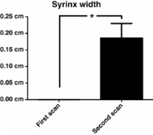
          Chiari-like malformation (CM) and syringomyelia is a neurological disease complex with high prevalence in cavalier King Charles spaniels (CKCS). The natural progression of this disease with time has not been described. The objectives of this study were to i) determine if syringomyelia progresses with time ii) determine if features of craniocrebral morphology previously associated with CM are progressive (including caudal cranial fossa volume, caudal cranial fossa parenchymal volume, ventricular dimensions, height of the foramen magnum and degree of cerebellar herniation). A retrospective morphometric analysis was undertaken in 12 CKCS with CM for which repeat magnetic resonance images were available without surgical intervention.

          Results

          The maximal syrinx width, height of the foramen magnum, length of cerebellar herniation and caudal cranial fossa volume increased over time. Ventricular and caudal fossa parenchymal volumes were not significantly different between scans.

          Conclusions

          The results of this study suggest that syringomyelia progresses with time. Increased caudal cranial fossa volume may be associated with active resorption of the supraoccipital bone, which has previously been found in histology specimens from adult CKCS. We hypothesise that active resorption of the supraoccipital bone occurs due to pressure from the cerebellum. These findings have important implications for our understanding of the pathogenesis and variable natural clinical progression of CM and syringomyelia in CKCS.

          Related collections

          Most cited references31

          • Record: found
          • Abstract: found
          • Article: not found

          Chiari I malformation redefined: clinical and radiographic findings for 364 symptomatic patients.

          Chiari malformations are regarded as a pathological continuum of hindbrain maldevelopments characterized by downward herniation of the cerebellar tonsils. The Chiari I malformation (CMI) is defined as tonsillar herniation of at least 3 to 5 mm below the foramen magnum. Increased detection of CMI has emphasized the need for more information regarding the clinical features of the disorder. We examined a prospective cohort of 364 symptomatic patients. All patients underwent magnetic resonance imaging of the head and spine, and some were evaluated using CINE-magnetic resonance imaging and other neurodiagnostic tests. For 50 patients and 50 age- and gender-matched control subjects, the volume of the posterior cranial fossa was calculated by the Cavalieri method. The families of 21 patients participated in a study of familial aggregation. There were 275 female and 89 male patients. The age of onset was 24.9+/-15.8 years (mean +/- standard deviation), and 89 patients (24%) cited trauma as the precipitating event. Common associated problems included syringomyelia (65%), scoliosis (42%), and basilar invagination (12%). Forty-three patients (12%) reported positive family histories of CMI or syringomyelia. Pedigrees for 21 families showed patterns consistent with autosomal dominant or recessive inheritance. The clinical syndrome of CMI was found to consist of the following: 1) headaches, 2) pseudotumor-like episodes, 3) a Meniere's disease-like syndrome, 4) lower cranial nerve signs, and 5) spinal cord disturbances in the absence of syringomyelia. The most consistent magnetic resonance imaging findings were obliteration of the retrocerebellar cerebrospinal fluid spaces (364 patients), tonsillar herniation of at least 5 mm (332 patients), and varying degrees of cranial base dysplasia. Volumetric calculations for the posterior cranial fossa revealed a significant reduction of total volume (mean, 13.4 ml) and a 40% reduction of cerebrospinal fluid volume (mean, 10.8 ml), with normal brain volume. These data support accumulating evidence that CMI is a disorder of the para-axial mesoderm that is characterized by underdevelopment of the posterior cranial fossa and overcrowding of the normally developed hindbrain. Tonsillar herniation of less than 5 mm does not exclude the diagnosis. Clinical manifestations of CMI seem to be related to cerebrospinal fluid disturbances (which are responsible for headaches, pseudotumor-like episodes, endolymphatic hydrops, syringomyelia, and hydrocephalus) and direct compression of nervous tissue. The demonstration of familial aggregation suggests a genetic component of transmission.
            Bookmark
            • Record: found
            • Abstract: found
            • Article: not found

            Development and tissue origins of the mammalian cranial base.

            The vertebrate cranial base is a complex structure composed of bone, cartilage and other connective tissues underlying the brain; it is intimately connected with development of the face and cranial vault. Despite its central importance in craniofacial development, morphogenesis and tissue origins of the cranial base have not been studied in detail in the mouse, an important model organism. We describe here the location and time of appearance of the cartilages of the chondrocranium. We also examine the tissue origins of the mouse cranial base using a neural crest cell lineage cell marker, Wnt1-Cre/R26R, and a mesoderm lineage cell marker, Mesp1-Cre/R26R. The chondrocranium develops between E11 and E16 in the mouse, beginning with development of the caudal (occipital) chondrocranium, followed by chondrogenesis rostrally to form the nasal capsule, and finally fusion of these two parts via the midline central stem and the lateral struts of the vault cartilages. X-Gal staining of transgenic mice from E8.0 to 10 days post-natal showed that neural crest cells contribute to all of the cartilages that form the ethmoid, presphenoid, and basisphenoid bones with the exception of the hypochiasmatic cartilages. The basioccipital bone and non-squamous parts of the temporal bones are mesoderm derived. Therefore the prechordal head is mostly composed of neural crest-derived tissues, as predicted by the New Head Hypothesis. However, the anterior location of the mesoderm-derived hypochiasmatic cartilages, which are closely linked with the extra-ocular muscles, suggests that some tissues associated with the visual apparatus may have evolved independently of the rest of the "New Head".
              Bookmark
              • Record: found
              • Abstract: found
              • Article: not found

              Syringomyelia: current concepts in pathogenesis, diagnosis, and treatment.

              Syringomyelia is a condition that results in fluid-containing cavities within the parenchyma of the spinal cord as a consequence of altered cerebrospinal fluid dynamics. This review discusses the history and the classification of the disorder, the current theories of pathogenesis, and the advanced imaging modalities used in the diagnosis. The intramedullary pulse pressure theory (a new pathophysiologic concept of syringomyelia) also is presented. In addition, the current understanding of the painful nature of this condition is discussed and the current trends in medical and surgical management are reviewed.
                Bookmark

                Author and article information

                Journal
                BMC Vet Res
                BMC Vet. Res
                BMC Veterinary Research
                BioMed Central
                1746-6148
                2012
                8 November 2012
                : 8
                : 215
                Affiliations
                [1 ]Department of Veterinary Clinical Sciences, Royal Veterinary College, London, UK
                [2 ]2The Animal Health Trust, Newmarket, UK
                [3 ]Stone Lion Veterinary Hospital, Goddard Veterinary Group, London, UK
                [4 ]Depertment of Veterinary Basic Sciences, Royal Veterinary College, London, UK
                Article
                1746-6148-8-215
                10.1186/1746-6148-8-215
                3514376
                23136935
                448c0d8a-11cb-4514-a539-c1cba6a85522
                Copyright ©2012 Driver et al.; licensee BioMed Central Ltd.

                This is an Open Access article distributed under the terms of the Creative Commons Attribution License ( http://creativecommons.org/licenses/by/2.0), which permits unrestricted use, distribution, and reproduction in any medium, provided the original work is properly cited.

                History
                : 4 April 2012
                : 31 October 2012
                Categories
                Methodology Article

                Veterinary medicine
                Veterinary medicine

                Comments

                Comment on this article