13
views
0
recommends
+1 Recommend
0 collections
    0
    shares
      • Record: found
      • Abstract: found
      • Article: found
      Is Open Access

      Team Flow Is a Unique Brain State Associated with Enhanced Information Integration and Interbrain Synchrony

      research-article

      Read this article at
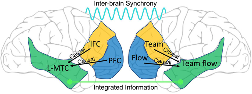
      Bookmark
          There is no author summary for this article yet. Authors can add summaries to their articles on ScienceOpen to make them more accessible to a non-specialist audience.

          Visual Abstract

          Abstract

          Team flow occurs when a group functions in a high task engagement to achieve a goal, commonly seen in performance and sports. Team flow can enable enhanced positive experiences, as compared with individual flow or regular socializing. However, the neural basis for this enhanced behavioral state remains unclear. Here, we identified neural correlates (NCs) of team flow in human participants using a music rhythm task with electroencephalogram hyperscanning. Experimental manipulations held the motor task constant while disrupting the corresponding hedonic music to interfere with the flow state or occluding the partner’s positive feedback to impede team interaction. We validated these manipulations by using psychometric ratings and an objective measure for the depth of flow experience, which uses the auditory-evoked potential (AEP) of a task-irrelevant stimulus. Spectral power analysis at both the scalp sensors and anatomic source levels revealed higher β-γ power specific to team flow in the left middle temporal cortex (L-MTC). Causal interaction analysis revealed that the L-MTC is downstream in information processing and receives information from areas encoding the flow or social states. The L-MTC significantly contributes to integrating information. Moreover, we found that team flow enhances global interbrain integrated information (II) and neural synchrony. We conclude that the NCs of team flow induce a distinct brain state. Our results suggest a neurocognitive mechanism to create this unique experience.

          Related collections

          Most cited references65

          • Record: found
          • Abstract: not found
          • Article: not found

          The use of fast Fourier transform for the estimation of power spectra: A method based on time averaging over short, modified periodograms

          P. Welch (1967)
            Bookmark
            • Record: found
            • Abstract: found
            • Article: found
            Is Open Access

            Brainstorm: A User-Friendly Application for MEG/EEG Analysis

            Brainstorm is a collaborative open-source application dedicated to magnetoencephalography (MEG) and electroencephalography (EEG) data visualization and processing, with an emphasis on cortical source estimation techniques and their integration with anatomical magnetic resonance imaging (MRI) data. The primary objective of the software is to connect MEG/EEG neuroscience investigators with both the best-established and cutting-edge methods through a simple and intuitive graphical user interface (GUI).
              Bookmark
              • Record: found
              • Abstract: found
              • Article: not found

              Automatic parcellation of human cortical gyri and sulci using standard anatomical nomenclature.

              Precise localization of sulco-gyral structures of the human cerebral cortex is important for the interpretation of morpho-functional data, but requires anatomical expertise and is time consuming because of the brain's geometric complexity. Software developed to automatically identify sulco-gyral structures has improved substantially as a result of techniques providing topologically correct reconstructions permitting inflated views of the human brain. Here we describe a complete parcellation of the cortical surface using standard internationally accepted nomenclature and criteria. This parcellation is available in the FreeSurfer package. First, a computer-assisted hand parcellation classified each vertex as sulcal or gyral, and these were then subparcellated into 74 labels per hemisphere. Twelve datasets were used to develop rules and algorithms (reported here) that produced labels consistent with anatomical rules as well as automated computational parcellation. The final parcellation was used to build an atlas for automatically labeling the whole cerebral cortex. This atlas was used to label an additional 12 datasets, which were found to have good concordance with manual labels. This paper presents a precisely defined method for automatically labeling the cortical surface in standard terminology. Copyright 2010 Elsevier Inc. All rights reserved.
                Bookmark

                Author and article information

                Journal
                eNeuro
                eNeuro
                eneuro
                eNeuro
                eNeuro
                Society for Neuroscience
                2373-2822
                4 October 2021
                12 October 2021
                Sep-Oct 2021
                : 8
                : 5
                : ENEURO.0133-21.2021
                Affiliations
                [1 ]Division of Biology and Biological Engineering, California Institute of Technology , Pasadena 91125, CA
                [2 ]The Electronics-Inspired Interdisciplinary Research Institute (EIIRIS), Toyohashi University of Technology , Toyohashi 441-8580, Japan
                [3 ]The University of Hong Kong , Pokfulam 999077, Hong Kong
                [4 ]NTT Communication Science Laboratories, NTT Corporation , Atsugi 243-0198, Japan
                [5 ]School of Psychological Sciences and Turner Institute for Brain and Mental Health, Monash University , Melbourne, Victoria 3800, Australia
                [6 ]Center for Information and Neural Networks (CiNet), National Institute of Information and Communications Technology (NICT) , Suita 565-0871, Japan
                [7 ]Advanced Telecommunications Research Computational Neuroscience Laboratories , Kyoto 619-0288, Japan
                [8 ]Research Institute of Electrical Communication, Tohoku University , Sendai 980-8577, Japan
                [9 ]Department of Computer Science and Engineering, Toyohashi University of Technology, Toyohashi 441-8580, Japan
                Author notes

                The authors declare no competing financial interests.

                Author contributions: M.S., M.C., and S.S. designed research; M.S., M.C., and D.-A.W. performed research; M.S., M.C., A.L., N.T., and S.S. analyzed data; M.S., M.C., A.L., N.T., D.-A.W., C.-h.T., S.N., and S.S. wrote the paper.

                This work was supported by the Program for Promoting the Enhancement of Research Universities funded to Toyohashi University of Technology and Grants-in-Aid for Scientific Research (Fostering Joint International Research(B), Grant Number 18KK0280) (M.S. and S.N.), Sponsored Research by Qneuro, Inc. (M.S. and S.S.), Translational Research Institute through NASA Cooperative Agreement NNX16AO69A (M.S. and S.S.), and by the Japan Science and Technology (JST)-CREST Grant JPMJCR14E4 (to S.S.). M.C. is supported by the University of Hong Kong Postgraduate Scholarship Program. C.-h.T. is supported by the University of Hong Kong General Research Fund and the Cooperative Research Project Program of the Research Institute of Electrical Communication, Tohoku University. N.T. is supported by Australian Research Council Discovery Projects Grants DP180104128 and DP180100396. A.L. is supported by an Australian Government Research Training Program Scholarship.

                Correspondence should be addressed to Mohammad Shehata at mohammad.shehata@ 123456gmail.com .
                Author information
                https://orcid.org/0000-0003-1710-3009
                https://orcid.org/0000-0003-0611-6170
                https://orcid.org/0000-0003-4216-8701
                https://orcid.org/0000-0003-4296-3369
                Article
                eN-NWR-0133-21
                10.1523/ENEURO.0133-21.2021
                8513532
                34607804
                448674d7-8132-4cd5-bb19-63532fe18d4d
                Copyright © 2021 Shehata et al.

                This is an open-access article distributed under the terms of the Creative Commons Attribution 4.0 International license, which permits unrestricted use, distribution and reproduction in any medium provided that the original work is properly attributed.

                History
                : 29 March 2021
                : 19 August 2021
                : 7 September 2021
                Page count
                Figures: 6, Tables: 1, Equations: 0, References: 66, Pages: 17, Words: 00
                Funding
                Funded by: Japan Society for the Promotion of Science (JSPS), doi 10.13039/501100001691;
                Award ID: JPMJCR14E4
                Funded by: Australian Research Council (ARC), doi 10.13039/501100000923;
                Award ID: DP180104128 and DP180100396
                Funded by: Toyohashi University of Technology (TUT), doi 10.13039/501100005984;
                Funded by: University of Hong Kong (HKU), doi 10.13039/501100003803;
                Funded by: Australian Government Research Training Program
                Categories
                1
                Research Article: New Research
                Cognition and Behavior
                Custom metadata
                September/October 2021

                eeg,flow,hyperscanning,in the zone,neural synchrony,teams
                eeg, flow, hyperscanning, in the zone, neural synchrony, teams

                Comments

                Comment on this article

                scite_
                0
                0
                0
                0
                Smart Citations
                0
                0
                0
                0
                Citing PublicationsSupportingMentioningContrasting
                View Citations

                See how this article has been cited at scite.ai

                scite shows how a scientific paper has been cited by providing the context of the citation, a classification describing whether it supports, mentions, or contrasts the cited claim, and a label indicating in which section the citation was made.

                Similar content42

                Cited by5

                Most referenced authors1,095