22
views
0
recommends
+1 Recommend
0 collections
    0
    shares
      • Record: found
      • Abstract: found
      • Article: not found

      Oxidative Stress and the Induction of Cyclooxygenase Enzymes and Apoptosis in the Murine Placenta

      research-article

      Read this article at

      ScienceOpenPublisherPMC
      Bookmark
          There is no author summary for this article yet. Authors can add summaries to their articles on ScienceOpen to make them more accessible to a non-specialist audience.

          Abstract

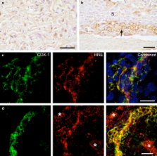
          Placental oxidative stress has been implicated in many complications of human pregnancy, including preterm delivery and preeclampsia. It is now appreciated that reactive oxygen species can induce a spectrum of changes, ranging from homeostatic induction of enzymes to apoptotic cell death. Little is known regarding the occurrence of placental oxidative stress in other species. We investigated markers of oxidative stress in the labyrinthine (LZ) and junctional (JZ) zones of the murine placenta across gestational age, and correlated these with expression of the cyclooxygenase enzymes COX-1 and COX-2, and apoptosis. We tested a causal link between the two by subjecting placental explants to hypoxia-reoxygenation (H/R) in vitro, a known stimulus for generation of oxidative stress. Western blotting demonstrated significant increases in the concentrations of hydroxynonenal (HNE), COX-1 and COX-2 with gestational age. Dual-labelling demonstrated co-localisation of HNE, and COX-1 and COX-2 within the trophoblast of the LZ, and glycogen cells of the JZ. An apoptotic index based on TUNEL-positivity demonstrated an increase with gestational age, and dual-labelling showed co-localisation of TUNEL labelling with HNE and active caspase-3 within the trophoblast of the LZ. H/R significantly increased oxidative stress, induction of COX-1 and COX-2, and the apoptotic index. Co-localisation demonstrated the increases in COX to be within the trophoblast of the LZ, and in particular the glycogen cells of the JZ. Apoptosis was restricted to the LZ. We speculate that the induction of COX enzymes is a physiological response to oxidative stress, and may play a role in initiating or augmenting parturition. Generation of oxidative stress may also play a role in influencing the growth trajectory of the placenta, and its component cell types. The mouse may provide an experimental genetic model in which to investigate these phenomena.

          Related collections

          Most cited references38

          • Record: found
          • Abstract: found
          • Article: not found

          Comparative developmental anatomy of the murine and human definitive placentae.

          The placenta of eutherian mammals is a remarkable biological structure. It is composed of both zygote-derived and maternal cells, and mediates the complex interactions between the mother and the fetus that are necessary for fetal growth and survival. While the genetic basis of human placental development and function is largely unknown, its understanding is of immense clinical importance because placentopathies of unknown genetic aetiology are thought to be the cause of many types of pregnancy complications including unexplained miscarriage and intrauterine growth retardation. The mouse is the best-studied mammalian experimental genetic model system and research is not restricted by the inherent ethical and practical limitations associated with the human. As a result, knowledge about the genetic control of mouse placental development has expanded greatly in recent years. In order for this to be of benefit to medical practice, extrapolations from murine to human placentation have to be made. However, comprehensive comparisons of the placentae of these two species are rare. This review therefore compares the developmental anatomy of the placenta between humans and mice with emphasis on structures and cell types that might be analogous between the two species. This could be of particular benefit to mouse developmental geneticists who study placental development and have an interest in the possible clinical implications of their work. Copyright 2002 Harcourt Publishers Ltd.
            Bookmark
            • Record: found
            • Abstract: found
            • Article: not found

            Immunocytochemical detection and mapping of a cytokeratin 18 neo-epitope exposed during early apoptosis.

            A neo-epitope in cytokeratin 18 (CK18) that becomes available at an early caspase cleavage event during apoptosis and is not detectable in vital epithelial cells is characterized. The monoclonal antibody M30, specific for this site, can be utilized specifically to recognize apoptotic cells, which show cytoplasmic cytokeratin filaments and aggregates after immunohistochemistry with M30, while viable and necrotic cells are negative. The number of cells recognized by the antibody increases after induction of apoptosis in exponentially growing epithelial cell lines and immunoreactivity is independent of the phosphorylation state of the cytokeratins. The generation of the M30 neo-epitope occurs early in the apoptotic cascade, before annexin V reactivity or positive DNA nick end labelling. In a flow cytometric assay, the majority of the M30-positive cells appear in the 'apoptotic' subG1 peak. Tests with synthetic peptides define positions 387-396 of CK18, with a liberated C-terminus at the caspase cleavage site DALD-S, as the ten-residue epitope of M30. This epitope starts at the end of coil 2 of the predicted CK18 structure, at a probable hinge region, compatible with the sensitivity to proteolytic cleavage. The definition of a specific caspase cleavage site in CK18 as a neo-epitope can be used for quantification of apoptotic epithelial cells with immunocytochemical techniques and is applicable to both fresh and formalin-fixed material. Copyright 1999 John Wiley & Sons, Ltd.
              Bookmark
              • Record: found
              • Abstract: found
              • Article: not found

              Oxidative stress in the pathogenesis of preeclampsia.

              The etiology and pathogenesis of the pregnancy syndrome preeclampsia remain poorly understood. There is substantial evidence to suggest that the diverse manifestations of preeclampsia, including altered vascular reactivity, vasospasm, and discrete pathology in many organ systems, are derived from pathologic changes within the maternal vascular endothelium. With the theme of endothelial cell dysfunction emphasized, this review focuses on the role of oxidative stress (an imbalance favoring oxidant over antioxidant forces) in the pathogenesis of preeclampsia. Data are summarized regarding 1) the role of the placenta in preeclampsia; 2) evidence and mechanisms of oxidative stress in the preeclampsia placenta; 3) markers of oxidative stress in the maternal circulation; and 4) the potential role of maternal dyslipidemia in generation of oxidative stress. A recurrent theme is that free radical reactions, promoted by "cross-talk" between the diseased placenta and maternal dyslipidemia, promote a vicious cycle of events that make cause and effect difficult to distinguish but likely contribute to the progression of preeclampsia.
                Bookmark

                Author and article information

                Journal
                Placenta
                Placenta
                W.B. Saunders
                0143-4004
                1532-3102
                July 2007
                July 2007
                : 28
                : 7
                : 724-733
                Affiliations
                Department of Physiology, Development and Neuroscience, University of Cambridge, Cambridge CB2 3DY, UK
                Author notes
                []Corresponding author. Present address: Physiological Laboratory, Downing Street, Cambridge CB2 3EG, UK. Tel.: +44 1223 333 856; fax: +44 1223 333 840. gjb2@ 123456cam.ac.uk
                [1]

                These authors contributed equally to the study.

                Article
                YPLAC1585
                10.1016/j.placenta.2006.12.001
                1895600
                17222904
                44584be2-27bf-4f31-a9e2-863f0dceb042
                .

                This document may be redistributed and reused, subject to certain conditions.

                History
                : 1 December 2006
                Categories
                Article

                Obstetrics & Gynecology
                cyclooxygenases,apoptosis,murine placenta,oxidative stress
                Obstetrics & Gynecology
                cyclooxygenases, apoptosis, murine placenta, oxidative stress

                Comments

                Comment on this article