8
views
0
recommends
+1 Recommend
0 collections
    0
    shares
      • Record: found
      • Abstract: found
      • Article: found
      Is Open Access

      Aging affects the biological activity of fibroblast growth factor (FGF) in gastric epithelial cell, which is partially rescued by uridine

      research-article
      a , a , a , a , a , b , b , a
      Bioengineered
      Taylor & Francis
      FGF, FGFR, aging, gastric epithelial cell, uridine

      Read this article at

      Bookmark
          There is no author summary for this article yet. Authors can add summaries to their articles on ScienceOpen to make them more accessible to a non-specialist audience.

          ABSTRACT

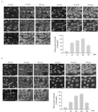
          Aging has become an irreversible trend in the world, the health problems caused by aging cannot be ignored. The physiological functions of human body begin to decline with aging, the decline of gastrointestinal function caused by aging is an important problem that needs to be resolved. In this work, we evaluated the anti-aging effect of uridine in the senescent gastric epithelial cell model, and found that the aging level of gastric epithelial cell was significantly down-regulated by uridine treatment, uridine could obviously down-regulate the ratio of the SA-β-gal-positive senescent cells. Furthermore, aging-related marker molecules (such as p16 and p21) were also significantly down-regulated under uridine treatment. Additionally, the levels of inflammation and oxidative stress were also significantly reduced by uridine treatment. Next, our further studies the effect of aging on FGF activity on gastric epithelial cell, and found that FGF/FGFR-mediated signaling pathways were significantly down-regulated. However, uridine treatment can not only alleviate the senescence of gastric epithelial cell, but also can partially restore the sensitivity of FGF signaling. Taken together, the current work indicates that uridine shows a good anti-aging effect, which lays a solid foundation for the related research in this filed.

          Graphical abstract

          Related collections

          Most cited references29

          • Record: found
          • Abstract: found
          • Article: not found

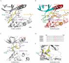
          Crystal structure of a ternary FGF-FGFR-heparin complex reveals a dual role for heparin in FGFR binding and dimerization.

          The crystal structure of a dimeric 2:2:2 FGF:FGFR:heparin ternary complex at 3 A resolution has been determined. Within each 1:1 FGF:FGFR complex, heparin makes numerous contacts with both FGF and FGFR, thereby augmenting FGF-FGFR binding. Heparin also interacts with FGFR in the adjoining 1:1 FGF:FGFR complex to promote FGFR dimerization. The 6-O-sulfate group of heparin plays a pivotal role in mediating both interactions. The unexpected stoichiometry of heparin binding in the structure led us to propose a revised model for FGFR dimerization. Biochemical data in support of this model are also presented. This model provides a structural basis for FGFR activation by small molecule heparin analogs and may facilitate the design of heparin mimetics capable of modulating FGF signaling.
            Bookmark
            • Record: found
            • Abstract: found
            • Article: found
            Is Open Access

            Fibroblast Growth Factor Receptors (FGFRs): Structures and Small Molecule Inhibitors

            Fibroblast growth factor receptors (FGFRs) are a family of receptor tyrosine kinases expressed on the cell membrane that play crucial roles in both developmental and adult cells. Dysregulation of FGFRs has been implicated in a wide variety of cancers, such as urothelial carcinoma, hepatocellular carcinoma, ovarian cancer and lung adenocarcinoma. Due to their functional importance, FGFRs have been considered as promising drug targets for the therapy of various cancers. Multiple small molecule inhibitors targeting this family of kinases have been developed, and some of them are in clinical trials. Furthermore, the pan-FGFR inhibitor erdafitinib (JNJ-42756493) has recently been approved by the U.S. Food and Drug Administration (FDA) for the treatment of metastatic or unresectable urothelial carcinoma (mUC). This review summarizes the structure of FGFR, especially its kinase domain, and the development of small molecule FGFR inhibitors.
              Bookmark
              • Record: found
              • Abstract: found
              • Article: not found

              Potentiation of glutathione loss and nerve cell death by the transition metals iron and copper: Implications for age-related neurodegenerative diseases

              There is growing evidence for alterations in iron and copper homeostasis during aging that are exacerbated in neurodegenerative diseases such as Alzheimer's disease (AD). However, how iron and copper accumulation leads to nerve cell damage in AD is not clear. In order to better understand how iron and copper can contribute to nerve cell death, a simple, well-defined in vitro model of cell death, the oyxtosis assay, was used. This assay uses glutamate to induce glutathione (GSH) depletion which initiates a form of oxidative stress-induced programmed cell death. A reduction in GSH is seen in the aging brain, is associated with cognitive dysfunction and is accelerated in many CNS diseases including AD. It is shown that both iron and copper potentiate both GSH loss and cell death in this model. Iron and copper also potentiate cell death induced by other GSH depleters but not by compounds that induce oxidative stress via other pathways. At least part of the effects of copper on GSH are related to its ability to reduce the activity of glutamate cysteine ligase, the rate limiting enzyme in GSH synthesis. Both metals also alter several signaling pathways involved in modulating nerve cell death. Together, these results suggest that in vivo iron and copper may specifically enhance nerve cell death under conditions where GSH levels are reduced.
                Bookmark

                Author and article information

                Journal
                Bioengineered
                Bioengineered
                Bioengineered
                Taylor & Francis
                2165-5979
                2165-5987
                1 February 2022
                2022
                1 February 2022
                : 13
                : 2
                : 3724-3738
                Affiliations
                [a ]School of Pharmacy, Jilin Medical University; , Jilin city, Jilin Province, 132013 China
                [b ]School of Pharmacy, Beihua University; , Jilin City, China
                Author notes
                CONTACT Xin Sun zxmjlmc@ 123456163.com Jilin Medical University;
                Article
                2029066
                10.1080/21655979.2022.2029066
                8974118
                35105283
                43d5f8a2-8beb-4c42-acd7-9f11871fdba8
                © 2022 The Author(s). Published by Informa UK Limited, trading as Taylor & Francis Group.

                This is an Open Access article distributed under the terms of the Creative Commons Attribution License ( http://creativecommons.org/licenses/by/4.0/), which permits unrestricted use, distribution, and reproduction in any medium, provided the original work is properly cited.

                History
                Page count
                Figures: 11, References: 29, Pages: 15
                Categories
                Research Article
                Research Paper

                Biomedical engineering
                fgf,fgfr,aging,gastric epithelial cell,uridine
                Biomedical engineering
                fgf, fgfr, aging, gastric epithelial cell, uridine

                Comments

                Comment on this article