16
views
0
recommends
+1 Recommend
0 collections
    0
    shares
      • Record: found
      • Abstract: found
      • Article: found
      Is Open Access

      Cytometry of chromatin bound Mcm6 and PCNA identifies two states in G1 that are separated functionally by the G1 restriction point 1

      research-article
      1 , 1 ,
      BMC Cell Biology
      BioMed Central

      Read this article at

      Bookmark
          There is no author summary for this article yet. Authors can add summaries to their articles on ScienceOpen to make them more accessible to a non-specialist audience.

          Abstract

          Background

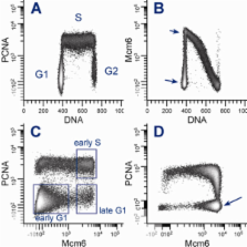
          Cytometric measurements of DNA content and chromatin-bound Mcm2 have demonstrated bimodal patterns of expression in G1. These patterns, the replication licensing function of Mcm proteins, and a correlation between Mcm loading and cell cycle commitment for cells re-entering the cell cycle, led us to test the idea that cells expressing a defined high level of chromatin-bound Mcm6 in G1 are committed - i.e., past the G1 restriction point. We developed a cell-based assay for tightly-bound PCNA (PCNA*) and Mcm6 (Mcm6*), DNA content, and a mitotic marker to clearly define G1, S, G2, and M phases of the cell cycle. hTERT-BJ1, hTERT-RPE-1, and Molt4 cells were extracted with Triton X-100 followed by methanol fixation, stained with antibodies and DAPI, then measured by cytometry.

          Results

          Bivariate analysis of cytometric data demonstrated complex patterns with distinct clustering for all combinations of the 4 variables. In G1, cells clustered in two groups characterized by low and high Mcm6* expression. Serum starvation and release experiments showed that residence in the high group was in late G1, just prior to S phase. Kinetic experiments, employing serum withdrawal, and stathmokinetic analysis with aphidicolin, mimosine or nocodazole demonstrated that cells with high levels of Mcm6* cycled with the committed phases of the cell cycle (S, G2, and M).

          Conclusions

          A multivariate assay for Mcm6*, PCNA*, DNA content, and a mitotic marker provides analysis capable of estimating the fraction of pre and post-restriction point G1 cells and supports the idea that there are at least two states in G1 defined by levels of chromatin bound Mcm proteins.

          Related collections

          Most cited references34

          • Record: found
          • Abstract: found
          • Article: not found

          Excess MCM proteins protect human cells from replicative stress by licensing backup origins of replication.

          The six main minichromosome maintenance proteins (Mcm2-7), which presumably constitute the core of the replicative DNA helicase, are present in chromatin in large excess relative to the number of active replication forks. To evaluate the relevance of this apparent surplus of Mcm2-7 complexes in human cells, their levels were down-regulated by using RNA interference. Interestingly, cells continued to proliferate for several days after the acute (>90%) reduction of Mcm2-7 concentration. However, they became hypersensitive to DNA replication stress, accumulated DNA lesions, and eventually activated a checkpoint response that prevented mitotic division. When this checkpoint was abrogated by the addition of caffeine, cells quickly lost viability, and their karyotypes revealed striking chromosomal aberrations. Single-molecule analyses revealed that cells with a reduced concentration of Mcm2-7 complexes display normal fork progression but have lost the potential to activate "dormant" origins that serve a backup function during DNA replication. Our data show that the chromatin-bound "excess" Mcm2-7 complexes play an important role in maintaining genomic integrity under conditions of replicative stress.
            Bookmark
            • Record: found
            • Abstract: found
            • Article: not found

            The Pezcoller lecture: cancer cell cycles revisited.

            C Sherr (2000)
            Genetic lesions that disable key regulators of G1 phase progression in mammalian cells are present in most human cancers. Mitogen-dependent, cyclin D-dependent kinases (cdk4 and cdk6) phosphorylate the retinoblastoma (Rb) tumor suppressor protein, helping to cancel its growth-inhibitory effects and enabling E2F transcription factors to activate genes required for entry into the DNA synthetic phase (S) of the cell division cycle. Among the E2F-responsive genes are cyclins E and A, which combine with and activate cdk2 to facilitate S phase entry and progression. Accumulation of cyclin D-dependent kinases during G1 phase sequesters cdk2 inhibitors of the Cip/Kip family, complementing the effects of the E2F transcriptional program by facilitating cyclin E-cdk2 activation at the G1-S transition. Disruption of "the Rb pathway" results from direct mutational inactivation of Rb function, by overexpression of cyclin D-dependent kinases, or through loss of p16(INK4a), an inhibitor of the cyclin D-dependent kinases. Reduction in levels of p27(Kip1) and increased expression of cyclin E also occur and carry a poor prognostic significance in many common forms of cancer. The ARF tumor suppressor, encoded by an alternative reading frame of the INK4a-ARF locus, senses "mitogenic current" flowing through the Rb pathway and is induced by abnormal growth promoting signals. By antagonizing Mdm2, a negative regulator of the p53 tumor suppressor, ARF triggers a p53-dependent transcriptional response that diverts incipient cancer cells to undergo growth arrest or apoptosis. Although ARF is not directly activated by signals that damage DNA, its loss not only dampens the p53 response to abnormal mitogenic signals but also renders tumor cells resistant to treatment by cytotoxic drugs and irradiation. Lesions in the p16--cyclin D-CDK4--Rb and ARF--Mdm2--p53 pathways occur so frequently in cancer, regardless of patient age or tumor type, that they appear to be part of the life history of most, if not all, cancer cells.
              Bookmark
              • Record: found
              • Abstract: found
              • Article: not found

              Genome-wide studies highlight indirect links between human replication origins and gene regulation.

              To get insights into the regulation of replication initiation, we systematically mapped replication origins along 1% of the human genome in HeLa cells. We identified 283 origins, 10 times more than previously known. Origin density is strongly correlated with genomic landscapes, with clusters of closely spaced origins in GC-rich regions and no origins in large GC-poor regions. Origin sequences are evolutionarily conserved, and half of them map within or near CpG islands. Most of the origins overlap transcriptional regulatory elements, providing further evidence of a connection with gene regulation. Moreover, we identify c-JUN and c-FOS as important regulators of origin selection. Half of the identified replication initiation sites do not have an open chromatin configuration, showing the absence of a direct link with gene regulation. Replication timing analyses coupled with our origin mapping suggest that a relatively strict origin-timing program regulates the replication of the human genome.
                Bookmark

                Author and article information

                Journal
                BMC Cell Biol
                BMC Cell Biology
                BioMed Central
                1471-2121
                2010
                16 April 2010
                : 11
                : 26
                Affiliations
                [1 ]Case Comprehensive Cancer Center, Case Western Reserve University, Cleveland, OH 44106, USA
                Article
                1471-2121-11-26
                10.1186/1471-2121-11-26
                2882901
                20398392
                432bfdd0-ed11-474f-9225-eaaeb0932ed4
                Copyright ©2010 Frisa and Jacobberger; licensee BioMed Central Ltd.

                This is an Open Access article distributed under the terms of the Creative Commons Attribution License ( http://creativecommons.org/licenses/by/2.0), which permits unrestricted use, distribution, and reproduction in any medium, provided the original work is properly cited.

                History
                : 1 September 2009
                : 16 April 2010
                Categories
                Research article

                Cell biology
                Cell biology

                Comments

                Comment on this article