6
views
0
recommends
+1 Recommend
0 collections
    0
    shares
      • Record: found
      • Abstract: found
      • Article: found
      Is Open Access

      Evaluating the effect of injecting triamcinolone acetonide in two sessions for preventing esophageal stricture after endoscopic submucosal dissection

      research-article

      Read this article at

      Bookmark
          There is no author summary for this article yet. Authors can add summaries to their articles on ScienceOpen to make them more accessible to a non-specialist audience.

          Abstract

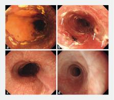
          Background and study aims  Several previous reports indicate that endoscopic injection of triamcinolone acetonide (TA) after widespread endoscopic submucosal dissection (ESD) is effective for preventing esophageal stricture. We investigated the efficacy of injecting TA in two sessions for preventing stricture formation post-ESD.

          Patients and methods  Sixty-six consecutive patients with widespread mucosal defects that affected more than three-fourths of the circumference of the esophagus were included. The study group (n = 40) received TA injections over two sessions: immediately after and 14 days after ESD. The control group (n = 26) did not receive a TA injection. This study was performed retrospectively against historical controls. The primary endpoint of this study was frequency of stricture after TA injection. The secondary endpoint was number of required endoscopic balloon dilations (EBDs) after TA injection.

          Results  The post-ESD stricture rate among patients who had subcircumferential mucosal defects was 45.7 % in the study group (16/35 patients), which was significantly lower than the rate of 73.9 % in the control group (17/23 patients; P  = 0.031). The number of EBD procedures required was significantly lower in the study group (median 0, range 0 – 7) than in the control group (median 4, range 0 – 20; P  < 0.001). There was no significant difference between the study and control groups among the patients who had full circumferential mucosal defects.

          Conclusion  This study showed that performing two sessions of TA injection is an effective and safe treatment for prevention of esophageal stricture following subcircumferential ESD.

          Related collections

          Most cited references21

          • Record: found
          • Abstract: found
          • Article: not found

          Predictors of postoperative stricture after esophageal endoscopic submucosal dissection for superficial squamous cell neoplasms.

          Although endoscopic submucosal dissection (ESD) is becoming accepted as an established treatment for superficial esophageal squamous cell neoplasms, the risks for developing postoperative stricture have not been elucidated. This was a retrospective study at a single institution. From January 2002 to October 2008, 65 patients with high-grade intraepithelial neoplasms (HGINs) or m2 carcinomas treated by ESD were enrolled. Predictors of postoperative stricture were investigated by comparing results from 11 patients who developed strictures with those from 54 patients who did not. Significant differences between the two groups were observed in longitudinal diameter (45.0 +/- 15.9 mm vs. 31.5 +/- 13.6 mm) and circumferential diameter (37.2 +/- 8.6 mm vs. 26.8 +/- 9.7 mm) of the resected specimens, and the proportion of extension to the whole circumference of the lumen ( 1 / 2/ > 3 / 4 : 2 / 4 / 5 vs. 40 / 13 / 1), histologic depth (HGIN/m2 : 2 / 9 vs. 41 / 13), and procedure time (85.6 +/- 42.8 minutes vs. 53.3 +/- 30.1 minutes). Multivariate analysis revealed that circumferential extension of > 3 / 4 (odds ration [OR]: 44.2; 95 % confidence interval [CI]: 4.4 - 443.6) and histologic depth to m2 (OR: 14.2; 95 %CI: 2.7 - 74.2) are reliable risk factors. Subanalysis for each category by combinations of these risk factors revealed that patients with lesions in > 3 / 4 of the circumferential area were associated with a high rate of postoperative stricture. By contrast, patients with HGIN lesions in < 3 / 4 extension have no probability of postoperative strictures. Additionally, subanalysis of patients with m2 lesions in < 3 / 4 circumferential extension revealed that circumferential diameter can be a reliable predictor for postoperative stricture. Circumferential extension and histologic depth are the reliable risk factors for postoperative strictures. In combination with circumferential diameter, we can perform effective and appropriate preventive balloon dilatations after esophageal ESD.
            Bookmark
            • Record: found
            • Abstract: found
            • Article: not found

            Endoscopic submucosal dissection of early esophageal cancer.

            In Japan, the majority of esophageal cancers are squamous cell carcinomas. Because no lymph node metastasis was reported in squamous cell carcinomas limited to the intraepithelial layer (m1) or proper mucosal layer (m2), the Japanese Esophageal Association recommended endoscopic mucosal resection (EMR) as the treatment of choice for these cancers. However, these lesions often spread laterally, exceeding the limits of en bloc resectability with conventional EMR methods such as the EMR cap method. The lesions resected in piece-meal manner with conventional EMR methods are prone to recur locally. Therefore, we developed a method of mucosal resection with a hook-knife that enables endoscopic submucosal dissection safely and achieves a high rate of en bloc resection for larger lesions. The median size of the resected specimen and cancer by our method was 32 mm (range, 8-76 mm) and 28 mm (range, 4-64 mm), respectively. The en bloc resection rate was 95% (95 of 102) and the local recurrence rate was 0% (0 of 102). This procedure was safe, with only 6 cases (6%) of mediastinal emphysema, which improved with conservative treatment. Endoscopic submucosal dissection with the hook knife is a method of endoluminal surgery enabling large en bloc resections without increased surgical risks.
              Bookmark
              • Record: found
              • Abstract: found
              • Article: not found

              The efficacy of endoscopic triamcinolone injection for the prevention of esophageal stricture after endoscopic submucosal dissection.

              Use of endoscopic submucosal dissection (ESD) for management of widespread superficial esophageal carcinomas may be complicated by the development of severe strictures, which may require serial treatment with endoscopic balloon dilatation (EBD). The goal of this study was to determine the efficacy of endoscopic triamcinolone injection (ETI) for the prevention of stricture formation after ESD. Case series. Tertiary-care referral center. A total of 41 consecutive patients who had a semi-circumferential mucosal defect that arose after ESD for superficial esophageal squamous cell carcinomas were enrolled in this study. EBD and ETI. Incidence of stricture and frequency of required EBD. ETI was performed in one group of patients (study group, n = 21) but not in the other (control group, n = 20). The incidence of stricture was significantly lower in the study group (19.0%) than in the control group (75.0%; P < .001). The number of required EBDs was also lower in the study group (mean, 1.7; range, 0-15) than in the control group (mean, 6.6; range 0-20). There were no side effects or complications associated with ETI. Nonrandomized study design and small number of patients in a single endoscopic center. This study suggests that ETI is safe and effective for the prevention of esophageal stricture in patients undergoing ESD for superficial esophageal squamous cell carcinomas. Copyright © 2011 American Society for Gastrointestinal Endoscopy. Published by Mosby, Inc. All rights reserved.
                Bookmark

                Author and article information

                Journal
                Endosc Int Open
                Endosc Int Open
                10.1055/s-00025476
                Endoscopy International Open
                © Georg Thieme Verlag KG (Stuttgart · New York )
                2364-3722
                2196-9736
                June 2019
                17 May 2019
                : 7
                : 6
                : E764-E770
                Affiliations
                [1 ]Niigata University Medical and Dental Hospital, Division of Gastroenterology and Hepatology, Niigata, Japan
                [2 ]Niigata University Medical and Dental Hospital, Department of Endoscopy, Niigata, Japan
                [3 ]Nagaoka Red Cross Hospital, Department of Gastroenterology, Niigata, Japan
                [4 ]Niigata Prefecture Yoshida Hospital, Department of Gastroenterology, Niigata, Japan
                [5 ]Niigata Cancer Center Hospital, Department of Gastroenterology, Niigata, Japan
                Author notes
                Corresponding author Satoru Hashimoto Division of Gastroenterology and Hepatology Niigata University Medical and Dental Hospital 1-757 Asahimachi-dori, Chuo-kuNiigata 951-8510Japan+81-25-227-0776 hashim@ 123456med.niigata-u.ac.jp
                Article
                10.1055/a-0894-4374
                6525005
                31157294
                42593ff4-e435-43d2-8bf3-c76ce1b5d6d0

                This is an open-access article distributed under the terms of the Creative Commons Attribution-NonCommercial-NoDerivatives License, which permits unrestricted reproduction and distribution, for non-commercial purposes only; and use and reproduction, but not distribution, of adapted material for non-commercial purposes only, provided the original work is properly cited.

                History
                : 23 September 2018
                : 14 March 2019
                Categories
                Original article

                Comments

                Comment on this article