701
views
0
recommends
+1 Recommend
0 collections
    0
    shares
      • Record: found
      • Abstract: found
      • Article: found
      Is Open Access

      Comparison of Laparoscopic and Microscopic Subinguinal Varicocelectomy in terms of Postoperative Scrotal Pain

      research-article

      Read this article at

      Bookmark
          There is no author summary for this article yet. Authors can add summaries to their articles on ScienceOpen to make them more accessible to a non-specialist audience.

          Abstract

          Laparoscopic varicocelectomy was found to be safe, effective, and minimally invasive. It appeared to result in less postoperative discomfort and earlier return to normal activity.

          Abstract

          Background and Objectives:

          In this study, 2 different varicocelectomy methods were compared with regard to postoperative scrotal pain, length of operation, and complications.

          Methods:

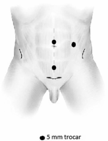
          Forty varicocele patients, who visited our clinic because of infertility or scrotal pain between 2008 and 2009, were enrolled in this clinical study. Microscopic subinguinal varicocelectomy was performed on 20 patients in Group I, and laparoscopic varicocelectomy was performed on 20 patients in Group II. Following surgery, the patients were assessed for postoperative requirements for analgesia; return to normal activity; varicocele recurrence; hydrocele formation; scrotal pain at postoperative days 1, 3, and 7; and other complications.

          Results:

          Mean age was 24.2±3.4 years in Group I and 25.1±2.1 years in Group II. Mean pain scores at postoperative 1, 3, and 7 days in Group I were (5.20±1.14, 4.60±0.97, and 3.50±0.97, respectively) significantly higher than those of Group II (0.70±0.82, 0.60±0.84, and 0.10±0.32, respectively). Time to return to normal activity was significantly shorter in Group II (3.7±2.1 days) compared with Group I (6.8±3.4 days) (p=0.028). However, the number of recurrences and hydroceles, as a complication of varicocelectomy, was 2 times higher in Group II (10%) than in Group I (5%).

          Conclusions:

          We believe that laparoscopic varicocelectomy is a safe, effective, and minimally invasive procedure. Furthermore, reduced postoperative discomfort and earlier return to normal activity are additional advantages of this method.

          Related collections

          Most cited references19

          • Record: found
          • Abstract: found
          • Article: not found

          Treatment of palpable varicocele in infertile men: a meta-analysis to define the best technique.

          To date, there have been no randomized, controlled, prospective clinical studies that compare various techniques to describe the best method for the treatment of varicocele in infertile men. This meta-analysis aims to address the best treatment modality for palpable varicocele in infertile men. A MEDLINE search was performed for articles published between January 1980 and April 2008, and we analyzed 36 studies reporting postoperative spontaneous pregnancy rates and/or complication rates after varicocele repair using various techniques in infertile men with palpable unilateral or bilateral varicocele. Spontaneous pregnancy rates and postoperative complications such as hydrocele formation, recurrence, or persistence were compared among the techniques. In addition, interventional failure with radiologic embolization and reported complications with the laparoscopic approach were reviewed. Overall spontaneous pregnancy rates were 37.69% in the Palomo technique series, 41.97% in the microsurgical varicocelectomy techniques, 30.07% in the laparoscopic varicocelectomy techniques, 33.2% in the radiologic embolization, and 36% in the macroscopic inguinal (Ivanissevich) varicocelectomy series, revealing significant differences among the techniques (P = .001). Overall recurrence rates were 14.97% in the Palomo technique series, 1.05% in the microsurgical varicocelectomy techniques, 4.3% in the laparoscopic varicocelectomy techniques, 12.7% in the radiologic embolization, and 2.63% in the macroscopic inguinal (Ivanissevich) or subinguinal varicocelectomy series, revealing significant difference among the techniques (P = .001). Overall hydrocele formation rates were 8.24% in the Palomo technique series, 0.44% in the microsurgical varicocelectomy techniques, 2.84% in the laparoscopic varicocelectomy, and 7.3% in the macroscopic inguinal (Ivanissevich) or subinguinal varicocelectomy series, revealing significant difference among the techniques (P = .001). We conclude that the microsurgical varicocelectomy technique has higher spontaneous pregnancy rates and lower postoperative recurrence and hydrocele formation than conventional varicocelectomy techniques in infertile men. However, prospective, randomized, and comparative studies with large number of patients are needed to compare the efficacy of microsurgical varicocelectomy with that of other treatment modalities in infertile men with varicocele.
            Bookmark
            • Record: found
            • Abstract: found
            • Article: not found

            Microsurgical inguinal varicocelectomy with delivery of the testis: an artery and lymphatic sparing technique.

            Conventional techniques of varicocele repair are associated with substantial risks of hydrocele formation, ligation of the testicular artery, and varicocele recurrence. We describe a microsurgical technique of varicocelectomy that significantly lowers the incidence of these complications. The testicle is delivered through a 2 to 3 cm. inguinal incision, and all external spermatic and gubernacular veins are ligated. The testis is returned to the scrotum and the spermatic cord is dissected under the operating microscope. The testicular artery and lymphatics are identified and preserved. All internal spermatic veins are doubly ligated with small hemoclips or 4-zero silk and divided. The vas deferens and its vessels are preserved. Initially, we performed 33 conventional inguinal varicocelectomies in 24 men without delivery of the testis or use of a microscope. Postoperatively, 3 unilateral hydroceles (9%) and 3 unilateral recurrences (9%) were detected. For the next 12 cases 2.5x loupes were used resulting in no hydroceles but another recurrence (8%). We then performed 640 varicocelectomies in 429 men using the microsurgical technique with delivery of the testis. Among 382 men available for followup examination from 6 months to 7 years postoperatively no hydroceles and no cases of testicular atrophy were found. A total of 4 unilateral recurrent varicoceles (0.6%) was identified. The differences between the techniques in the incidence of hydrocele formation and varicocele recurrence are highly significant (p < 0.001). No wound infections occurred in any men. Four scrotal hematomas (0.6%), 1 of which required surgical drainage, occurred in the group with microsurgical ligation and delivery of the testis compared to none with the conventional technique. Preoperative and postoperative semen analyses (mean 3.57 analyses per patient) were obtained on 271 men. The changes in sperm count x 10(6) cc (36.9 to 46.8, p < 0.001), per cent motility (39.6 to 45.7%, p < 0.001) and per cent normal forms (48.4 to 52.10%, p < 0.001) were highly significant. The pregnancy rate was 152 of 357 couples (43%) followed for a minimum of 6 months postoperatively. Delivery of the testis through a small inguinal incision provides direct visual access to all possible avenues of testicular venous drainage. The operating microscope allows identification of the testicular artery, lymphatics and small venous channels. This minimally invasive, outpatient technique results in a significant decrease in the incidence of hydrocele formation, testicular artery injury and varicocele recurrence.
              Bookmark
              • Record: found
              • Abstract: found
              • Article: not found

              Outcomes of varicocele ligation done for pain.

              Surgical ligation is an option in the management of patients with painful varicocele. Little objective data exist addressing the effectiveness of this treatment. We reviewed records from 58 patients who underwent varicocele ligation at our institution from January 1985 to May 1996 to establish success of surgical ligation of the painful varicocele. ICD-9 billing codes were used to identify all patients who had undergone varicocele ligation for pain since 1985. We documented patient age, grade and location of varicocele, duration and quality of pain, response to conservative therapy and surgical approach to ligation. Telephone interviews and chart reviews were conducted to determine resolution of pain, complications of the procedure and if the patient would choose surgery again. We obtained followup on 35 of the 58 painful varicocele patients (60%). Average patient age was 25.7 years (range 15 to 65). The varicocele was on the left side in 30 men and bilateral in 5. Of the patients 31 described the pain as a dull throbbing ache, 2 as sharp and 2 as a pulling sensation. Initial conservative therapy failed in all 35 men. Varicocele was grade III in 18 cases, grade II in 16 and grade I in 1. The inguinal or subinguinal approach was used in 24 patients, high ligation in 10 and laparoscopic repair in 1. In 30 patients there was (86%) complete resolution of pain postoperatively and 1 had partial resolution. Only 4 patients (11%) had persistent or worse symptoms. This retrospective review supports the conclusion that varicocele ligation is an effective treatment for painful varicocele in properly selected patients.
                Bookmark

                Author and article information

                Contributors
                Dicle University, Medical Faculty, Department of Urology, Diyarbakir, Turkey.
                Dicle University, Medical Faculty, Department of Urology, Diyarbakir, Turkey.
                Dicle University, Medical Faculty, Department of Urology, Diyarbakir, Turkey.
                Dicle University, Medical Faculty, Department of Urology, Diyarbakir, Turkey.
                Dicle University, Medical Faculty, Department of Urology, Diyarbakir, Turkey.
                Sutcu Imam University, Medical Faculty, Department of Urology, Kahramanmaras, Turkey.
                Journal
                JSLS
                JSLS
                jsls
                jsls
                JSLS
                JSLS : Journal of the Society of Laparoendoscopic Surgeons
                Society of Laparoendoscopic Surgeons (Miami, FL )
                1086-8089
                1938-3797
                Apr-Jun 2012
                : 16
                : 2
                : 212-217
                Affiliations
                Dicle University, Medical Faculty, Department of Urology, Diyarbakir, Turkey.
                Dicle University, Medical Faculty, Department of Urology, Diyarbakir, Turkey.
                Dicle University, Medical Faculty, Department of Urology, Diyarbakir, Turkey.
                Dicle University, Medical Faculty, Department of Urology, Diyarbakir, Turkey.
                Dicle University, Medical Faculty, Department of Urology, Diyarbakir, Turkey.
                Sutcu Imam University, Medical Faculty, Department of Urology, Kahramanmaras, Turkey.
                Author notes
                Address correspondence to: Assist. Prof. Dr. Haluk Söylemez, MD, Dicle University, Medical Faculty, Department of Urology, Diyarbakir, Turkey. Telephone: +90 412 248 8583, Pbx: +90 412 248 8440, E-mail: drhaluks@ 123456yahoo.com
                Article
                11-07-114
                10.4293/108680812X13427982376220
                3481232
                23477168
                4233b68a-4ff8-4e25-960b-5bd2207fe75f
                © 2012 by JSLS, Journal of the Society of Laparoendoscopic Surgeons.

                This is an Open Access article distributed under the terms of the Creative Commons Attribution Non-Commercial No Derivatives License ( http://creativecommons.org/licenses/by-nc-nd/3.0/), which permits for noncommercial use, distribution, and reproduction in any medium, provided the original work is properly cited and is not altered in any way.

                History
                Categories
                Scientific Papers

                Surgery
                varicocele,laparoscopy,microscopy,postoperative pain
                Surgery
                varicocele, laparoscopy, microscopy, postoperative pain

                Comments

                Comment on this article