7
views
0
recommends
+1 Recommend
0 collections
    0
    shares
      • Record: found
      • Abstract: found
      • Article: found
      Is Open Access

      Hypermethylation of the CTRP9 promoter region promotes Hcy induced VSMC lipid deposition and foam cell formation via negatively regulating ER stress

      research-article

      Read this article at

      Bookmark
          There is no author summary for this article yet. Authors can add summaries to their articles on ScienceOpen to make them more accessible to a non-specialist audience.

          Abstract

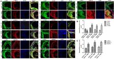
          To provide a theoretical basis for the prevention and treatment of atherosclerosis (As), the current study aimed to investigate the mechanism underlying the effect of homocysteine (Hcy) on inducing the lipid deposition and foam cell formation of the vascular smooth muscle cell (VSMC) via C1q/Tumor necrosis factor-related protein9 (CTRP9) promoter region Hypermethylation negative regulating endoplasmic reticulum stress (ERs). Therefore, apolipoprotein E deficient (ApoE −/−) mice were randomly divided into the control [ApoE −/− + normal diet (NC)] and high methionine [ApoE −/− + (normal diet supplemented with 1.7% methionine (HMD)] groups (n = 6 mice/group). Following feeding for 15 weeks, the serum levels of Homocysteine (Hcy), total cholesterol (TC), and triglyceride (TG) were measured using an automatic biochemical analyzer. HE and oil red O staining were performed on the aorta roots to observe the pathological changes. Additionally, immunofluorescence staining was performed to detect the protein expression levels of CTRP9, glucose-regulated protein 78 kD (GRP78), phosphorylated protein kinase RNA-like ER kinase (p-PERK), activating transcription factor 6a (ATF6a), phosphorylated inositol-requiring enzyme-1α (p-IRE1α), sterol regulatory element binding proteins-1c (SREBP1c) and sterol regulatory element binding proteins-2 (SREBP2) in VSMC derived from murine aortic roots. In vitro, VSMC was stimulated with 100 μmol/l Hcy. After transfection of plasmids with overexpression and interference of CTRP9, ERs agonist (TM) and inhibitor (4-PBA) were given to stimulate VSMC cells. HE staining and oil red O staining were used to observe the effect of Hcy stimulation on lipid deposition in VSMC. Additionally, The mRNA and protein expression levels of CTRP9, GRP78, PERK, ATF6a, IRE1α, SREBP1c, and SREBP2 in VSMC were detected by RT-qPCR and western blot analysis, respectively. Finally, The methylation modification of the CTRP9 promoter region has been studied. The NCBI database was used to search the promoter region of the CTRP9 gene, and CpG Island was used to predict the methylation site. After Hcy stimulation of VSMC, overexpression of DNMT1, and intervention with 5-Azc, assess the methylation level of the CTRP9 promoter through bisulfite sequencing PCR (BSP). The results showed that the serum levels of Hcy, TC, and TG in the ApoE −/− + HMD group were significantly increased compared with the ApoE −/− + NC group. In addition, HE staining and oil red O staining showed obvious AS plaque formation in the vessel wall, and a large amount of fat deposition in VSMC, thus indicating that the hyperhomocysteinemia As an animal model was successfully established. Furthermore, CTRP9 were downregulated, while GRP78, p-PERK, ATF6a, p-IRE1α, SREBP1c, SREBP2 was upregulated in aortic VSMC in the ApoE −/− + HMD group. Consistent with the in vivo results, Hcy can inhibit the expression of CTRP9 in VSMC and induce ERs and lipid deposition in VSMC. Meanwhile, the increased expression of CTRP9 can reduce ERs and protect the lipid deposition in Hcy induced VSMC. Furthermore, ERs can promote Hcy induced VSMC lipid deposition, inhibition of ERs can reduce Hcy induced VSMC lipid deposition, and CTRP9 may play a protective role in Hcy induced VSMC lipid deposition and foam cell transformation through negative regulation of ERs. In addition, The CTRP9 promoter in the Hcy group showed hypermethylation. At the same time as Hcy intervention, overexpression of DNMT1 increases the methylation level of the CTRP9 promoter, while 5-Azc can reduce the methylation level of the CTRP9 promoter. Finally, Hcy can up-regulate the expression of DNMT1 and down-regulate the expression of CTRP9. After overexpression of DNMT1, the expression of CTRP9 is further decreased. After 5-Azc inhibition of DNMT1, the expression of DNMT1 decreases, while the expression of CTRP9 increases. It is suggested that the molecular mechanism of Hcy inhibiting the expression of CTRP9 is related to the hypermethylation of the CTRP9 promoter induced by Hcy and regulated by DNMT1. 5-Azc can inhibit the expression of DNMT1 and reverse the regulatory effect of DNMT1 on CTRP9. Overall, the results of the present study suggested that Hcy induces DNA hypermethylation in the CTRP9 promoter region by up-regulating DNMT1 expression, and negatively regulates ERs mediated VSMC lipid deposition and foam cell formation. CTRP9 may potentially be a therapeutic target in the treatment of hyperhomocysteinemia and As.

          Related collections

          Most cited references32

          • Record: found
          • Abstract: found
          • Article: not found

          Analysis of relative gene expression data using real-time quantitative PCR and the 2(-Delta Delta C(T)) Method.

          The two most commonly used methods to analyze data from real-time, quantitative PCR experiments are absolute quantification and relative quantification. Absolute quantification determines the input copy number, usually by relating the PCR signal to a standard curve. Relative quantification relates the PCR signal of the target transcript in a treatment group to that of another sample such as an untreated control. The 2(-Delta Delta C(T)) method is a convenient way to analyze the relative changes in gene expression from real-time quantitative PCR experiments. The purpose of this report is to present the derivation, assumptions, and applications of the 2(-Delta Delta C(T)) method. In addition, we present the derivation and applications of two variations of the 2(-Delta Delta C(T)) method that may be useful in the analysis of real-time, quantitative PCR data. Copyright 2001 Elsevier Science (USA).
            Bookmark
            • Record: found
            • Abstract: found
            • Article: not found

            DNA methylation and its basic function.

            In the mammalian genome, DNA methylation is an epigenetic mechanism involving the transfer of a methyl group onto the C5 position of the cytosine to form 5-methylcytosine. DNA methylation regulates gene expression by recruiting proteins involved in gene repression or by inhibiting the binding of transcription factor(s) to DNA. During development, the pattern of DNA methylation in the genome changes as a result of a dynamic process involving both de novo DNA methylation and demethylation. As a consequence, differentiated cells develop a stable and unique DNA methylation pattern that regulates tissue-specific gene transcription. In this chapter, we will review the process of DNA methylation and demethylation in the nervous system. We will describe the DNA (de)methylation machinery and its association with other epigenetic mechanisms such as histone modifications and noncoding RNAs. Intriguingly, postmitotic neurons still express DNA methyltransferases and components involved in DNA demethylation. Moreover, neuronal activity can modulate their pattern of DNA methylation in response to physiological and environmental stimuli. The precise regulation of DNA methylation is essential for normal cognitive function. Indeed, when DNA methylation is altered as a result of developmental mutations or environmental risk factors, such as drug exposure and neural injury, mental impairment is a common side effect. The investigation into DNA methylation continues to show a rich and complex picture about epigenetic gene regulation in the central nervous system and provides possible therapeutic targets for the treatment of neuropsychiatric disorders.
              Bookmark
              • Record: found
              • Abstract: found
              • Article: not found

              Vascular smooth muscle cells in atherosclerosis

              Vascular smooth muscle cells (VSMCs) are a major cell type present at all stages of an atherosclerotic plaque. According to the 'response to injury' and 'vulnerable plaque' hypotheses, contractile VSMCs recruited from the media undergo phenotypic conversion to proliferative synthetic cells that generate extracellular matrix to form the fibrous cap and hence stabilize plaques. However, lineage-tracing studies have highlighted flaws in the interpretation of former studies, revealing that these studies had underestimated both the content and functions of VSMCs in plaques and have thus challenged our view on the role of VSMCs in atherosclerosis. VSMCs are more plastic than previously recognized and can adopt alternative phenotypes, including phenotypes resembling foam cells, macrophages, mesenchymal stem cells and osteochondrogenic cells, which could contribute both positively and negatively to disease progression. In this Review, we present the evidence for VSMC plasticity and summarize the roles of VSMCs and VSMC-derived cells in atherosclerotic plaque development and progression. Correct attribution and spatiotemporal resolution of clinically beneficial and detrimental processes will underpin the success of any therapeutic intervention aimed at VSMCs and their derivatives.
                Bookmark

                Author and article information

                Contributors
                zhangmh270@163.com
                Journal
                Sci Rep
                Sci Rep
                Scientific Reports
                Nature Publishing Group UK (London )
                2045-2322
                9 November 2023
                9 November 2023
                2023
                : 13
                : 19438
                Affiliations
                [1 ]Department of Pathophysiology, School of Basic Medical Sciences, Ningxia Medical University, ( https://ror.org/02h8a1848) 1160 Shengli Street, Yinchuan, 750004 Ningxia People’s Republic of China
                [2 ]Ningxia Key Laboratory of Vascular Injury and Repair Research, Key Laboratory of Metabolic Cardiovascular Diseases Research of National Health Commission, Yinchuan, 750004 Ningxia People’s Republic of China
                Article
                46981
                10.1038/s41598-023-46981-5
                10636064
                37945738
                4108e33f-27ee-4552-a469-f67e35baf6cb
                © The Author(s) 2023

                Open Access This article is licensed under a Creative Commons Attribution 4.0 International License, which permits use, sharing, adaptation, distribution and reproduction in any medium or format, as long as you give appropriate credit to the original author(s) and the source, provide a link to the Creative Commons licence, and indicate if changes were made. The images or other third party material in this article are included in the article's Creative Commons licence, unless indicated otherwise in a credit line to the material. If material is not included in the article's Creative Commons licence and your intended use is not permitted by statutory regulation or exceeds the permitted use, you will need to obtain permission directly from the copyright holder. To view a copy of this licence, visit http://creativecommons.org/licenses/by/4.0/.

                History
                : 29 May 2023
                : 7 November 2023
                Funding
                Funded by: Key Research and Development Project of Ningxia Hui Autonomous Region Natural Science Foundation
                Award ID: 2021BEG03093
                Funded by: Research Fund Project of Ningxia Medical University
                Award ID: XT2022028
                Categories
                Article
                Custom metadata
                © Springer Nature Limited 2023

                Uncategorized
                diseases,medical research,pathogenesis
                Uncategorized
                diseases, medical research, pathogenesis

                Comments

                Comment on this article