18
views
0
recommends
+1 Recommend
0 collections
    0
    shares
      • Record: found
      • Abstract: found
      • Article: found
      Is Open Access

      Attenuated Increase in Maximal Force of Rat Medial Gastrocnemius Muscle after Concurrent Peak Power and Endurance Training

      research-article

      Read this article at

      Bookmark
          There is no author summary for this article yet. Authors can add summaries to their articles on ScienceOpen to make them more accessible to a non-specialist audience.

          Abstract

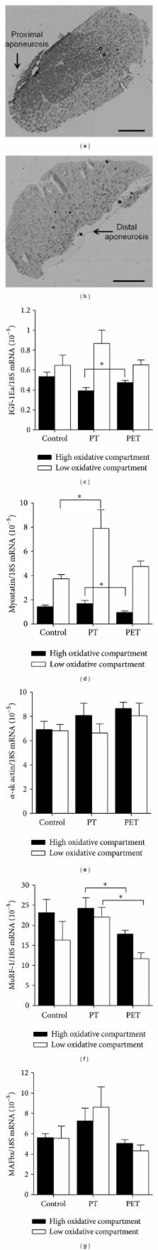
          Improvement of muscle peak power and oxidative capacity are generally presumed to be mutually exclusive. However, this may not be valid by using fibre type-specific recruitment. Since rat medial gastrocnemius muscle (GM) is composed of high and low oxidative compartments which are recruited task specifically, we hypothesised that the adaptive responses to peak power training were unaffected by additional endurance training. Thirty rats were subjected to either no training (control), peak power training (PT), or both peak power and endurance training (PET), which was performed on a treadmill 5 days per week for 6 weeks. Maximal running velocity increased 13.5% throughout the training and was similar in both training groups. Only after PT, GM maximal force was 10% higher than that of the control group. In the low oxidative compartment, mRNA levels of myostatin and MuRF-1 were higher after PT as compared to those of control and PET groups, respectively. Phospho-S6 ribosomal protein levels remained unchanged, suggesting that the elevated myostatin levels after PT did not inhibit mTOR signalling. In conclusion, even by using task-specific recruitment of the compartmentalized rat GM, additional endurance training interfered with the adaptive response of peak power training and attenuated the increase in maximal force after power training.

          Related collections

          Most cited references38

          • Record: found
          • Abstract: found
          • Article: not found

          Skeletal muscle hypertrophy and atrophy signaling pathways.

          Skeletal muscle hypertrophy is defined as an increase in muscle mass, which in the adult animal comes as a result of an increase in the size, as opposed to the number, of pre-existing skeletal muscle fibers. The protein growth factor insulin-like growth factor 1 (IGF-1) has been demonstrated to be sufficient to induce skeletal muscle hypertrophy. Over the past few years, signaling pathways which are activated by IGF-1, and which are responsible for regulating protein synthesis pathways, have been defined. More recently, it has been show that IGF-1 can also block the transcriptional upregulation of key mediators of skeletal muscle atrophy, the ubiquitin-ligases MuRF1 and MAFbx (also called Atrogin-1). Further, it has been demonstrated recently that activation of the NF-kappaB transcription pathway, activated by cachectic factors such as TNFalpha, is sufficient to induce skeletal muscle atrophy, and this atrophy occurs in part via NF-kappaB-mediated upregulation of MuRF1. Further work has demonstrated a trigger for MAFbx expression upon treatment with TNFalpha--the p38 MAPK pathway. This review will focus on the recent progress in the understanding of molecular signalling, which governs skeletal muscle atrophy and hypertrophy, and the known instances of cross-regulation between the two systems.
            Bookmark
            • Record: found
            • Abstract: found
            • Article: not found

            Myostatin reduces Akt/TORC1/p70S6K signaling, inhibiting myoblast differentiation and myotube size.

            Myostatin is a negative regulator of skeletal muscle size, previously shown to inhibit muscle cell differentiation. Myostatin requires both Smad2 and Smad3 downstream of the activin receptor II (ActRII)/activin receptor-like kinase (ALK) receptor complex. Other transforming growth factor-beta (TGF-beta)-like molecules can also block differentiation, including TGF-beta(1), growth differentiation factor 11 (GDF-11), activins, bone morphogenetic protein 2 (BMP-2) and BMP-7. Myostatin inhibits activation of the Akt/mammalian target of rapamycin (mTOR)/p70S6 protein synthesis pathway, which mediates both differentiation in myoblasts and hypertrophy in myotubes. Blockade of the Akt/mTOR pathway, using small interfering RNA to regulatory-associated protein of mTOR (RAPTOR), a component of TOR signaling complex 1 (TORC1), increases myostatin-induced phosphorylation of Smad2, establishing a myostatin signaling-amplification role for blockade of Akt. Blockade of RAPTOR also facilitates myostatin's inhibition of muscle differentiation. Inhibition of TORC2, via rapamycin-insensitive companion of mTOR (RICTOR), is sufficient to inhibit differentiation on its own. Furthermore, myostatin decreases the diameter of postdifferentiated myotubes. However, rather than causing upregulation of the E3 ubiquitin ligases muscle RING-finger 1 (MuRF1) and muscle atrophy F-box (MAFbx), previously shown to mediate skeletal muscle atrophy, myostatin decreases expression of these atrophy markers in differentiated myotubes, as well as other genes normally upregulated during differentiation. These findings demonstrate that myostatin signaling acts by blocking genes induced during differentiation, even in a myotube, as opposed to activating the distinct "atrophy program." In vivo, inhibition of myostatin increases muscle creatine kinase activity, coincident with an increase in muscle size, demonstrating that this in vitro differentiation measure is also upregulated in vivo.
              Bookmark
              • Record: found
              • Abstract: found
              • Article: not found

              The adaptations to strength training : morphological and neurological contributions to increased strength.

              High-resistance strength training (HRST) is one of the most widely practiced forms of physical activity, which is used to enhance athletic performance, augment musculo-skeletal health and alter body aesthetics. Chronic exposure to this type of activity produces marked increases in muscular strength, which are attributed to a range of neurological and morphological adaptations. This review assesses the evidence for these adaptations, their interplay and contribution to enhanced strength and the methodologies employed. The primary morphological adaptations involve an increase in the cross-sectional area of the whole muscle and individual muscle fibres, which is due to an increase in myofibrillar size and number. Satellite cells are activated in the very early stages of training; their proliferation and later fusion with existing fibres appears to be intimately involved in the hypertrophy response. Other possible morphological adaptations include hyperplasia, changes in fibre type, muscle architecture, myofilament density and the structure of connective tissue and tendons. Indirect evidence for neurological adaptations, which encompasses learning and coordination, comes from the specificity of the training adaptation, transfer of unilateral training to the contralateral limb and imagined contractions. The apparent rise in whole-muscle specific tension has been primarily used as evidence for neurological adaptations; however, morphological factors (e.g. preferential hypertrophy of type 2 fibres, increased angle of fibre pennation, increase in radiological density) are also likely to contribute to this phenomenon. Changes in inter-muscular coordination appear critical. Adaptations in agonist muscle activation, as assessed by electromyography, tetanic stimulation and the twitch interpolation technique, suggest small, but significant increases. Enhanced firing frequency and spinal reflexes most likely explain this improvement, although there is contrary evidence suggesting no change in cortical or corticospinal excitability. The gains in strength with HRST are undoubtedly due to a wide combination of neurological and morphological factors. Whilst the neurological factors may make their greatest contribution during the early stages of a training programme, hypertrophic processes also commence at the onset of training.
                Bookmark

                Author and article information

                Journal
                Biomed Res Int
                BMRI
                BioMed Research International
                Hindawi Publishing Corporation
                2314-6133
                2314-6141
                2013
                27 January 2013
                : 2013
                : 935671
                Affiliations
                1MOVE Research Institute Amsterdam, Faculty of Human Movement Sciences, VU University Amsterdam, Van der Boechorststraat 9, 1081 BT Amsterdam, The Netherlands
                2Department of Clinical Chemistry, MOVE Research Institute Amsterdam, VU University Medical Center Amsterdam, 1007 MB Amsterdam, The Netherlands
                3Department of Endocrinology, MOVE Research Institute Amsterdam, VU University Medical Center Amsterdam, 1007 MB Amsterdam, The Netherlands
                4Institute for Biomedical Research into Human Movement and Health, Manchester Metropolitan University, Manchester M5 1GD, UK
                Author notes
                *Regula Furrer: r.furrer@ 123456vu.nl

                Academic Editor: Mouldy Sioud

                Article
                10.1155/2013/935671
                3581157
                23509812
                3e8f69f5-2725-451b-bafc-1522a0f2c158
                Copyright © 2013 Regula Furrer et al.

                This is an open access article distributed under the Creative Commons Attribution License, which permits unrestricted use, distribution, and reproduction in any medium, provided the original work is properly cited.

                History
                : 2 October 2012
                : 21 November 2012
                Categories
                Research Article

                Comments

                Comment on this article