7
views
0
recommends
+1 Recommend
0 collections
    0
    shares
      • Record: found
      • Abstract: found
      • Article: found
      Is Open Access

      Wide Anterior Maxillary Reconstruction with Equine Bone Xenograft: A Case Report of 24-Month Follow-Up

      case-report

      Read this article at

      Bookmark
          There is no author summary for this article yet. Authors can add summaries to their articles on ScienceOpen to make them more accessible to a non-specialist audience.

          Abstract

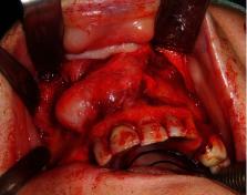
          Introduction. Orofacial reconstruction plays an important role in the treatment of patients affected by oral and maxillofacial cancers. Improvements in technologies and studies of biomaterials have widely expanded surgical possibilities to achieve good functional and aesthetic outcomes. By the way, xenografting procedures gained great consensus in the last decades, because of their documented reliability and efficacy. We present a case of anterior maxillary chondrosarcoma (CHS) that has undergone surgical ablation followed by reconstruction with an equine-derived bone xenograft. Case Presentation. A 68-year-old woman affected by CHS of the premaxilla underwent surgical ablation involving the four incisors followed by reconstruction using an equine-derived bone substitute. Bony reconstruction was planned to achieve implant and dental prosthetic rehabilitation at a second surgical time. Primary surgery was carried out without complications. Good integration of the graft was confirmed by radiological examination. At 12-month follow-up, the patient refused the implant placement and spontaneously adopted a mobile prosthesis. One year later, plates and screws were removed, because of the exposure of a titanium plate. The graft was finally rejected within 3 weeks. Discussion. Nonantigenic equine-derived biomaterials have shown reliability and a good safety profile. In the presented case, implant insertion should have been performed 12 months after the primary surgery. During the follow-up, until dental mobile prosthesis was applied, clinical and instrumental examinations demonstrated a good integration of the graft. We suppose that a chronic inflammation of the mucosa led to the exposure of the plate, perhaps due to pressure, minimal movements, or imperfect fitting of the mobile prosthesis. Removal of fixation means was performed to prevent grafting failure, without success. On the other hand, missing load could induce the graft to act just like a prosthesis, without a real process of integration. Safety and reliability of equine-derived bone xenografts cannot be currently confirmed if not followed by implant insertion and dental rehabilitation.

          Related collections

          Most cited references35

          • Record: found
          • Abstract: found
          • Article: found
          Is Open Access

          Bone regeneration: current concepts and future directions

          Bone regeneration is a complex, well-orchestrated physiological process of bone formation, which can be seen during normal fracture healing, and is involved in continuous remodelling throughout adult life. However, there are complex clinical conditions in which bone regeneration is required in large quantity, such as for skeletal reconstruction of large bone defects created by trauma, infection, tumour resection and skeletal abnormalities, or cases in which the regenerative process is compromised, including avascular necrosis, atrophic non-unions and osteoporosis. Currently, there is a plethora of different strategies to augment the impaired or 'insufficient' bone-regeneration process, including the 'gold standard' autologous bone graft, free fibula vascularised graft, allograft implantation, and use of growth factors, osteoconductive scaffolds, osteoprogenitor cells and distraction osteogenesis. Improved 'local' strategies in terms of tissue engineering and gene therapy, or even 'systemic' enhancement of bone repair, are under intense investigation, in an effort to overcome the limitations of the current methods, to produce bone-graft substitutes with biomechanical properties that are as identical to normal bone as possible, to accelerate the overall regeneration process, or even to address systemic conditions, such as skeletal disorders and osteoporosis.
            Bookmark
            • Record: found
            • Abstract: found
            • Article: not found
            Is Open Access

            β-tricalcium phosphate for bone substitution: Synthesis and properties

            β-tricalcium phosphate (β-TCP) is one the most used and potent synthetic bone graft substitute. It is not only osteoconductive, but also osteoinductive. These properties, combined with its cell-mediated resorption, allow full bone defects regeneration. Its clinical outcome is sometimes considered to be "unpredictable", possibly due to a poor understanding of β-TCP physico-chemical properties: β-TCP crystallographic structure is not fully uncovered; recent results suggest that sintered β-TCP is coated with a Ca-rich alkaline phase; β-TCP apatite-forming ability and osteoinductivity may be enhanced by a hydrothermal treatment; β-TCP grain size and porosity are strongly modified by the presence of minute amounts of β-calcium pyrophosphate or hydroxyapatite impurities. The aim of the present article is to provide a critical, but still rather comprehensive review of the current state of knowledge on β-TCP, with a strong focus on its synthesis and physico-chemical properties, and their link to the in vivo response. STATEMENT OF SIGNIFICANCE: The present review documents the richness, breadth, and interest of the research devoted to β-tricalcium phosphate (β-TCP). β-TCP is synthetic, osteoconductive, osteoinductive, and its resorption is cell-mediated, thus making it one of the most potent bone graft substitutes. This comprehensive review reveals that there are a number of aspects, such as surface chemistry, crystallography, or stoichiometry deviations, that are still poorly understood. As such, β-TCP is still an exciting scientific playground despite a 50 year long history and > 200 yearly publications.
              Bookmark
              • Record: found
              • Abstract: found
              • Article: not found

              Reconstruction of the maxilla and midface: introducing a new classification.

              Most patients requiring midface reconstruction have had ablative surgery for malignant disease, and most require postoperative radiotherapy. This type of facial reconstruction attracts controversy, not only because of the many reconstructive options, but also because dental and facial prostheses can be very successful in selected cases. This Personal View is based on a new classification of the midface defect, which emphasises the increasing complexity of the problem. Low defects not involving the orbital adnexae can often be successfully treated with dental obturators. For the more extensive maxillary defects, there is consensus that a free flap is required. Composite flaps of bone and muscle harvested from the iliac crest with internal oblique or the scapula tip with latissimus dorsi can more reliably support the orbit and cheek than soft-tissue free flaps and non-vascularised grafts, and also enable an implant-borne dental or orbital prosthesis. Nasomaxillary defects usually require bone to augment the loss of the nasal bones, but orbitomaxillary cases can be managed more simply with local or soft-tissue free flaps. We review the current options and our own experience over the past 15 years in an attempt to rationalise the management of these defects.
                Bookmark

                Author and article information

                Contributors
                Journal
                Case Rep Surg
                Case Rep Surg
                CRIS
                Case Reports in Surgery
                Hindawi
                2090-6900
                2090-6919
                2020
                21 October 2020
                : 2020
                : 8890935
                Affiliations
                1Maxillofacial Surgery, University of Naples “Federico II”, Naples, Italy
                2Maxillofacial Surgery Unit, University of Campania “Luigi Vanvitelli”, Naples, Italy
                3Dermatology, University of Insubria, Varese, Italy
                Author notes

                Academic Editor: Mario Ganau

                Author information
                https://orcid.org/0000-0003-4363-388X
                https://orcid.org/0000-0003-2177-9677
                https://orcid.org/0000-0002-8289-5527
                https://orcid.org/0000-0002-4497-3563
                Article
                10.1155/2020/8890935
                7596435
                3e8ebfe8-7a57-4bf1-a3bb-476381729ca0
                Copyright © 2020 Davide De Cicco et al.

                This is an open access article distributed under the Creative Commons Attribution License, which permits unrestricted use, distribution, and reproduction in any medium, provided the original work is properly cited.

                History
                : 10 April 2020
                : 8 October 2020
                : 13 October 2020
                Categories
                Case Report

                Surgery
                Surgery

                Comments

                Comment on this article