218
views
0
recommends
+1 Recommend
0 collections
    1
    shares
      • Record: found
      • Abstract: found
      • Article: found
      Is Open Access

      Structural and Functional Analysis of Human HtrA3 Protease and Its Subdomains

      research-article

      Read this article at

      Bookmark
          There is no author summary for this article yet. Authors can add summaries to their articles on ScienceOpen to make them more accessible to a non-specialist audience.

          Abstract

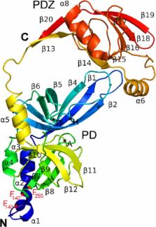
          Human HtrA3 protease, which induces mitochondria-mediated apoptosis, can be a tumor suppressor and a potential therapeutic target in the treatment of cancer. However, there is little information about its structure and biochemical properties. HtrA3 is composed of an N-terminal domain not required for proteolytic activity, a central serine protease domain and a C-terminal PDZ domain. HtrA3S, its short natural isoform, lacks the PDZ domain which is substituted by a stretch of 7 C-terminal amino acid residues, unique for this isoform. This paper presents the crystal structure of the HtrA3 protease domain together with the PDZ domain (ΔN-HtrA3), showing that the protein forms a trimer whose protease domains are similar to those of human HtrA1 and HtrA2. The ΔN-HtrA3 PDZ domains are placed in a position intermediate between that in the flat saucer-like HtrA1 SAXS structure and the compact pyramidal HtrA2 X-ray structure. The PDZ domain interacts closely with the LB loop of the protease domain in a way not found in other human HtrAs. ΔN-HtrA3 with the PDZ removed (ΔN-HtrA3-ΔPDZ) and an N-terminally truncated HtrA3S (ΔN-HtrA3S) were fully active at a wide range of temperatures and their substrate affinity was not impaired. This indicates that the PDZ domain is dispensable for HtrA3 activity. As determined by size exclusion chromatography, ΔN-HtrA3 formed stable trimers while both ΔN-HtrA3-ΔPDZ and ΔN-HtrA3S were monomeric. This suggests that the presence of the PDZ domain, unlike in HtrA1 and HtrA2, influences HtrA3 trimer formation. The unique C-terminal sequence of ΔN-HtrA3S appeared to have little effect on activity and oligomerization. Additionally, we examined the cleavage specificity of ΔN-HtrA3. Results reported in this paper provide new insights into the structure and function of ΔN-HtrA3, which seems to have a unique combination of features among human HtrA proteases.

          Related collections

          Most cited references43

          • Record: found
          • Abstract: found
          • Article: not found

          Structural basis of substrate specificity in the serine proteases.

          Structure-based mutational analysis of serine protease specificity has produced a large database of information useful in addressing biological function and in establishing a basis for targeted design efforts. Critical issues examined include the function of water molecules in providing strength and specificity of binding, the extent to which binding subsites are interdependent, and the roles of polypeptide chain flexibility and distal structural elements in contributing to specificity profiles. The studies also provide a foundation for exploring why specificity modification can be either straightforward or complex, depending on the particular system.
            Bookmark
            • Record: found
            • Abstract: found
            • Article: not found

            OMP peptide signals initiate the envelope-stress response by activating DegS protease via relief of inhibition mediated by its PDZ domain.

            Transmembrane signaling between intracellular compartments is often controlled by regulated proteolysis. Escherichia coli respond to misfolded or unfolded outer-membrane porins (OMPs) in the periplasm by inducing sigma(E)-dependent transcription of stress genes in the cytoplasm. This process requires a proteolytic cascade initiated by the DegS protease, which destroys a transmembrane protein (RseA) that normally binds to and inhibits sigma(E). Here, we show that peptides ending with OMP-like C-terminal sequences bind the DegS PDZ domain, activate DegS cleavage of RseA, and induce sigma(E)-dependent transcription. These results suggest that DegS acts as a sensor of envelope stress by binding unassembled OMPs. DegS activation involves relief of inhibitory interactions between its PDZ and protease domains. Peptide binding to inhibitory PDZ domains in proteases related to DegS, including DegP/HtrA, may also regulate the degradation of specific substrates by these enzymes.
              Bookmark
              • Record: found
              • Abstract: found
              • Article: not found

              Crystal structure of DegP (HtrA) reveals a new protease-chaperone machine.

              Molecular chaperones and proteases monitor the folded state of other proteins. In addition to recognizing non-native conformations, these quality control factors distinguish substrates that can be refolded from those that need to be degraded. To investigate the molecular basis of this process, we have solved the crystal structure of DegP (also known as HtrA), a widely conserved heat shock protein that combines refolding and proteolytic activities. The DegP hexamer is formed by staggered association of trimeric rings. The proteolytic sites are located in a central cavity that is only accessible laterally. The mobile side-walls are constructed by twelve PDZ domains, which mediate the opening and closing of the particle and probably the initial binding of substrate. The inner cavity is lined by several hydrophobic patches that may act as docking sites for unfolded polypeptides. In the chaperone conformation, the protease domain of DegP exists in an inactive state, in which substrate binding in addition to catalysis is abolished.
                Bookmark

                Author and article information

                Contributors
                Role: Editor
                Journal
                PLoS One
                PLoS ONE
                plos
                plosone
                PLoS ONE
                Public Library of Science (San Francisco, CA USA )
                1932-6203
                25 June 2015
                2015
                : 10
                : 6
                : e0131142
                Affiliations
                [1 ]Department of Biochemistry, Faculty of Biology, University of Gdansk, 80–308 Gdansk, Poland
                [2 ]Midwest Center for Structural Genomics, Argonne National Laboratory, Argonne, Illinois, IL 60439, United States of America
                [3 ]Structural Biology Center, Biosciences Division, Argonne National Laboratory, Argonne, Illinois, IL 60439, United States of America
                [4 ]Department of Biochemistry, Faculty of Chemistry, University of Gdansk, 80–308 Gdansk, Poland
                [5 ]Department of Molecular and Cellular Biology, Intercollegiate Faculty of Biotechnology of the University of Gdansk and the Medical University of Gdansk, 80–822 Gdansk, Poland
                Centro Nacional de Biotecnologia - CSIC, SPAIN
                Author notes

                Competing Interests: The authors have declared that no competing interests exist.

                Conceived and designed the experiments: BL JO AJ AL DZ-J BB. Performed the experiments: PG JO TW MJ AL BB. Analyzed the data: BL JO PG TW AL JS-G BB. Wrote the paper: BL JO DZ-J.

                Article
                PONE-D-15-06420
                10.1371/journal.pone.0131142
                4481513
                26110759
                3d7ea485-6e34-454b-a064-166f9d6512f7
                Copyright @ 2015

                This is an open access article distributed under the terms of the Creative Commons Attribution License, which permits unrestricted use, distribution, and reproduction in any medium, provided the original author and source are credited

                History
                : 11 February 2015
                : 27 May 2015
                Page count
                Figures: 11, Tables: 6, Pages: 24
                Funding
                This work was supported by National Institutes of Health Grant number GM094585 to A.J. and by the National Science Center (Poland), project grant no. UMO-2013/09/B/NZ1/01068 to B.L. Argonne is operated by UChicago Argonne, LLC, for the U.S. Department of Energy, Office of Biological and Environmental Research under contract DE-AC02-06CH11357. The funders had no role in study design, data collection and analysis, decision to publish, or preparation of the manuscript.
                Categories
                Research Article
                Custom metadata
                All relevant data are within the paper and in the Protein Data Bank database (accession number 4RI0).

                Uncategorized
                Uncategorized

                Comments

                Comment on this article