19
views
0
recommends
+1 Recommend
0 collections
    0
    shares
      • Record: found
      • Abstract: found
      • Article: found
      Is Open Access

      Localized Electrical Impedance Myography of the Biceps Brachii Muscle during Different Levels of Isometric Contraction and Fatigue

      research-article

      Read this article at

      Bookmark
          There is no author summary for this article yet. Authors can add summaries to their articles on ScienceOpen to make them more accessible to a non-specialist audience.

          Abstract

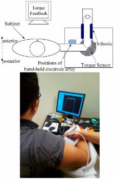
          This study assessed changes in electrical impedance myography (EIM) at different levels of isometric muscle contraction as well as during exhaustive exercise at 60% maximum voluntary contraction (MVC) until task failure. The EIM was performed on the biceps brachii muscle of 19 healthy subjects. The results showed that there was a significant difference between the muscle resistance (R) measured during the isometric contraction and when the muscle was completely relaxed. Post hoc analysis shows that the resistance increased at higher contractions (both 60% MVC and MVC), however, there were no significant changes in muscle reactance (X) during the isometric contractions. The resistance also changed during different stages of the fatigue task and there were significant decreases from the beginning of the contraction to task failure as well as between task failure and post fatigue rest. Although our results demonstrated an increase in resistance during isometric contraction, the changes were within 10% of the baseline value. These changes might be related to the modest alterations in muscle architecture during a contraction. The decrease in resistance seen with muscle fatigue may be explained by an accumulation of metabolites in the muscle tissue.

          Related collections

          Most cited references38

          • Record: found
          • Abstract: found
          • Article: not found

          A comparison of central aspects of fatigue in submaximal and maximal voluntary contractions.

          Magnetic and electrical stimulation at different levels of the neuraxis show that supraspinal and spinal factors limit force production in maximal isometric efforts ("central fatigue"). In sustained maximal contractions, motoneurons become less responsive to synaptic input and descending drive becomes suboptimal. Exercise-induced activity in group III and IV muscle afferents acts supraspinally to limit motor cortical output but does not alter motor cortical responses to transcranial magnetic stimulation. "Central" and "peripheral" fatigue develop more slowly during submaximal exercise. In sustained submaximal contractions, central fatigue occurs in brief maximal efforts even with a weak ongoing contraction (<15% maximum). The presence of central fatigue when much of the available motor pathway is not engaged suggests that afferent inputs contribute to reduce voluntary activation. Small-diameter muscle afferents are likely to be activated by local activity even in sustained weak contractions. During such contractions, it is difficult to measure central fatigue, which is best demonstrated in maximal efforts. To show central fatigue in submaximal contractions, changes in motor unit firing and force output need to be characterized simultaneously. Increasing central drive recruits new motor units, but the way this occurs is likely to depend on properties of the motoneurons and the inputs they receive in the task. It is unclear whether such factors impair force production for a set level of descending drive and thus represent central fatigue. The best indication that central fatigue is important during submaximal tasks is the disproportionate increase in subjects' perceived effort when maintaining a low target force.
            Bookmark
            • Record: found
            • Abstract: found
            • Article: not found

            Electrical impedance myography: Background, current state, and future directions.

            Electrical impedance myography (EIM) is a non-invasive technique for the evaluation of neuromuscular disease that relies upon the application and measurement of high-frequency, low-intensity electrical current. EIM assesses disease-induced changes to the normal composition and architecture of muscle, including myocyte atrophy and loss, edema, reinnervation, and deposition of endomysial connective tissue and fat. With application of single-frequency electrical current, EIM can be used to help grade the severity of neuromuscular disease. Assessing electrical impedance across a spectrum of applied frequencies and with current flow at multiple orientations relative to major muscle fiber direction can provide a more complete picture of the condition of muscle. EIM holds the promise of serving as an indicator of disease status. It may be useful in clinical trials and in monitoring effectiveness of treatment in individual patients; ultimately, it may also find diagnostic application. Ongoing efforts have been focused on obtaining a deeper understanding of the basic mechanisms of impedance change, studying EIM in a variety of clinical contexts, and further refining the methods of EIM data acquisition and analysis.
              Bookmark
              • Record: found
              • Abstract: found
              • Article: not found

              Localized bioimpedance analysis in the evaluation of neuromuscular disease.

              Localized bioimpedance analysis is a novel, noninvasive technique with potential application to neuromuscular disease. In this procedure, high-frequency alternating current is passed through muscle, and parameters related to the consequent voltage pattern are evaluated. Currents flowing perpendicular to muscle fibers encounter many more cell membranes than do currents flowing parallel to them, producing surface voltage patterns that are altered by disease. Using this technique, 45 normal subjects and 25 patients with various neuromuscular diseases were studied, including 4 with amyotrophic lateral sclerosis, 4 with inflammatory myopathy, and 11 with inclusion-body myositis. Two parameters, the spatially averaged phase and the effective longitudinal resistivity, were altered in patients with neuromuscular disease. Reductions in phase correlated to disease progression, whereas normalization of phase correlated with disease remission. In patients with inclusion-body myositis, a unique pattern of reduced phase and elevated resistivity was identified. These findings suggest that localized bioimpedance analysis has the potential of playing a substantial role in the diagnostic and therapeutic evaluation of neuromuscular disease. Copyright 2002 Wiley Periodicals, Inc.
                Bookmark

                Author and article information

                Contributors
                Role: Academic Editor
                Role: Academic Editor
                Journal
                Sensors (Basel)
                Sensors (Basel)
                sensors
                Sensors (Basel, Switzerland)
                MDPI
                1424-8220
                22 April 2016
                April 2016
                : 16
                : 4
                : 581
                Affiliations
                [1 ]Department of Rehabilitation Medicine, the First Affiliated Hospital, Sun Yat-sen University, Guangzhou 510080, China; Le.Li@ 123456uth.tmc.edu
                [2 ]Department of Physical Medicine and Rehabilitation, University of Texas Health Science Center at Houston; TIRR Memorial Hermann Research Center, Houston, TX 77030, USA; Henry.Shin@ 123456uth.tmc.edu (H.S.); Xiaoyan.Li@ 123456uth.tmc.edu (X.L.); Sheng.Li@ 123456uth.tmc.edu (S.L.)
                [3 ]Guangdong Work Injury Rehabilitation Center, Guangzhou 510440, China
                Author notes
                [* ]Correspondence: Ping.Zhou.1@ 123456uth.tmc.edu ; Tel.: +1-713-797-7126
                Article
                sensors-16-00581
                10.3390/s16040581
                4851095
                27110795
                3d740296-49cf-4b2e-aace-3e4f32be095e
                © 2016 by the authors; licensee MDPI, Basel, Switzerland.

                This article is an open access article distributed under the terms and conditions of the Creative Commons Attribution (CC-BY) license ( http://creativecommons.org/licenses/by/4.0/).

                History
                : 08 February 2016
                : 17 April 2016
                Categories
                Article

                Biomedical engineering
                electrical impedance myography,muscle contraction,fatigue
                Biomedical engineering
                electrical impedance myography, muscle contraction, fatigue

                Comments

                Comment on this article