24
views
0
recommends
+1 Recommend
0 collections
    0
    shares
      • Record: found
      • Abstract: found
      • Article: found
      Is Open Access

      Independent Component Analysis of the Effect of L-dopa on fMRI of Language Processing

      research-article

      Read this article at

      Bookmark
          There is no author summary for this article yet. Authors can add summaries to their articles on ScienceOpen to make them more accessible to a non-specialist audience.

          Abstract

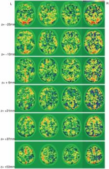
          L-dopa, which is a precursor for dopamine, acts to amplify strong signals, and dampen weak signals as suggested by previous studies. The effect of L-dopa has been demonstrated in language studies, suggesting restriction of the semantic network. In this study, we aimed to examine the effect of L-dopa on language processing with fMRI using Independent Component Analysis (ICA). Two types of language tasks (phonological and semantic categorization tasks) were tested under two drug conditions (placebo and L-dopa) in 16 healthy subjects. Probabilistic ICA (PICA), part of FSL, was implemented to generate Independent Components (IC) for each subject for the four conditions and the ICs were classified into task-relevant source groups by a correlation threshold criterion. Our key findings include: (i) The highly task-relevant brain regions including the Left Inferior Frontal Gyrus (LIFG), Left Fusiform Gyrus (LFUS), Left Parietal lobe (LPAR) and Superior Temporal Gyrus (STG) were activated with both L-dopa and placebo for both tasks, and (ii) as compared to placebo, L-dopa was associated with increased activity in posterior regions, including the superior temporal area (BA 22), and decreased activity in the thalamus (pulvinar) and inferior frontal gyrus (BA 11/47) for both tasks. These results raise the possibility that L-dopa may exert an indirect effect on posterior regions mediated by the thalamus (pulvinar).

          Related collections

          Most cited references30

          • Record: found
          • Abstract: found
          • Article: not found

          Role of left inferior prefrontal cortex in retrieval of semantic knowledge: a reevaluation.

          A number of neuroimaging findings have been interpreted as evidence that the left inferior frontal gyrus (IFG) subserves retrieval of semantic knowledge. We provide a fundamentally different interpretation, that it is not retrieval of semantic knowledge per se that is associated with left IFG activity but rather selection of information among competing alternatives from semantic memory. Selection demands were varied across three semantic tasks in a single group of subjects. Functional magnetic resonance imaging signal in overlapping regions of left IFG was dependent on selection demands in all three tasks. In addition, the degree of semantic processing was varied independently of selection demands in one of the tasks. The absence of left IFG activity for this comparison counters the argument that the effects of selection can be attributed solely to variations in degree of semantic retrieval. Our findings suggest that it is selection, not retrieval, of semantic knowledge that drives activity in the left IFG.
            Bookmark
            • Record: found
            • Abstract: found
            • Article: not found

            Analysis of fMRI data by blind separation into independent spatial components.

            Current analytical techniques applied to functional magnetic resonance imaging (fMRI) data require a priori knowledge or specific assumptions about the time courses of processes contributing to the measured signals. Here we describe a new method for analyzing fMRI data based on the independent component analysis (ICA) algorithm of Bell and Sejnowski ([1995]: Neural Comput 7:1129-1159). We decomposed eight fMRI data sets from 4 normal subjects performing Stroop color-naming, the Brown and Peterson work/number task, and control tasks into spatially independent components. Each component consisted of voxel values at fixed three-dimensional locations (a component "map"), and a unique associated time course of activation. Given data from 144 time points collected during a 6-min trial, ICA extracted an equal number of spatially independent components. In all eight trials, ICA derived one and only one component with a time course closely matching the time course of 40-sec alternations between experimental and control tasks. The regions of maximum activity in these consistently task-related components generally overlapped active regions detected by standard correlational analysis, but included frontal regions not detected by correlation. Time courses of other ICA components were transiently task-related, quasiperiodic, or slowly varying. By utilizing higher-order statistics to enforce successively stricter criteria for spatial independence between component maps, both the ICA algorithm and a related fourth-order decomposition technique (Comon [1994]: Signal Processing 36:11-20) were superior to principal component analysis (PCA) in determining the spatial and temporal extent of task-related activation. For each subject, the time courses and active regions of the task-related ICA components were consistent across trials and were robust to the addition of simulated noise. Simulated movement artifact and simulated task-related activations added to actual fMRI data were clearly separated by the algorithm. ICA can be used to distinguish between nontask-related signal components, movements, and other artifacts, as well as consistently or transiently task-related fMRI activations, based on only weak assumptions about their spatial distributions and without a priori assumptions about their time courses. ICA appears to be a highly promising method for the analysis of fMRI data from normal and clinical populations, especially for uncovering unpredictable transient patterns of brain activity associated with performance of psychomotor tasks.
              Bookmark
              • Record: found
              • Abstract: found
              • Article: not found

              Dopaminergic modulation of cognitive function-implications for L-DOPA treatment in Parkinson's disease.

              It is well recognised that patients with Parkinson's disease exhibit cognitive deficits, even in the earliest disease stages. Whereas, L-DOPA therapy in early Parkinson's disease is accepted to improve the motor symptoms, the effects on cognitive performance are more complex: both positive and negative effects have been observed. The purpose of the present article is to review the effects of L-DOPA medication in Parkinson's disease on cognitive functions in the broad domains of cognitive flexibility and working memory. The review places the effects in Parkinson's disease within a framework of evidence from studies with healthy human volunteers, rodents and non-human primates as well as computational modeling work. It is suggested that beneficial or detrimental effects of L-DOPA are observed depending on task demands and basal dopamine levels in distinct parts of the striatum. The study of the beneficial and detrimental cognitive effects of L-DOPA in Parkinson's disease has substantial implications for the understanding and treatment development of cognitive abnormalities in Parkinson's disease as well as normal health.
                Bookmark

                Author and article information

                Contributors
                Role: Editor
                Journal
                PLoS One
                plos
                plosone
                PLoS ONE
                Public Library of Science (San Francisco, USA )
                1932-6203
                2010
                17 August 2010
                : 5
                : 8
                : e11933
                Affiliations
                [1 ]Department of Statistics, The Ohio State University, Columbus, Ohio, United States of America
                [2 ]Department of Imaging Science and the Rochester Center for Brain Imaging, University of Rochester, Rochester, New York, United States of America
                [3 ]Department of Psychology, University of Massachusetts-Lowell, Lowell, Massachusetts, United States of America
                [4 ]Departments of Radiology, Neurology and Psychology, University of Missouri, Columbia, Missouri, United States of America
                [5 ]The Thompson Center, University of Missouri, Columbia, Missouri, United States of America
                Cuban Neuroscience Center, Cuba
                Author notes

                Conceived and designed the experiments: MET AH DQB. Performed the experiments: MET AH DQB. Analyzed the data: NK PG MET DQB. Contributed reagents/materials/analysis tools: NK PG. Wrote the paper: NK PG DQB.

                Article
                10-PONE-RA-17782
                10.1371/journal.pone.0011933
                2923146
                20808963
                3d3bddca-fe9d-4824-8c00-d7da19ebadea
                Kim et al. This is an open-access article distributed under the terms of the Creative Commons Attribution License, which permits unrestricted use, distribution, and reproduction in any medium, provided the original author and source are credited.
                History
                : 8 April 2010
                : 26 May 2010
                Page count
                Pages: 10
                Categories
                Research Article
                Pharmacology
                Neuroscience/Cognitive Neuroscience
                Physiology/Cognitive Neuroscience
                Radiology and Medical Imaging/Magnetic Resonance Imaging

                Uncategorized
                Uncategorized

                Comments

                Comment on this article