28
views
0
recommends
+1 Recommend
0 collections
    0
    shares
      • Record: found
      • Abstract: found
      • Article: found
      Is Open Access

      Mechanosensitive EPLIN-dependent remodeling of adherens junctions regulates epithelial reshaping

      research-article

      Read this article at

      Bookmark
          There is no author summary for this article yet. Authors can add summaries to their articles on ScienceOpen to make them more accessible to a non-specialist audience.

          Abstract

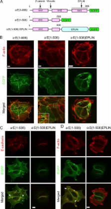
          The F-actin–stabilizing protein EPLIN is a mechanosensitive regulator of adherens junction remodeling in epithelial cells.

          Abstract

          The zonula adherens (ZA), a type of adherens junction (AJ), plays a major role in epithelial cell–cell adhesions. It remains unknown how the ZA is remodeled during epithelial reorganization. Here we found that the ZA was converted to another type of AJ with punctate morphology (pAJ) at the margins of epithelial colonies. The F-actin–stabilizing protein EPLIN (epithelial protein lost in neoplasm), which functions to maintain the ZA via its association with αE-catenin, was lost in the pAJs. Consistently, a fusion of αE-catenin and EPLIN contributed to the formation of ZA but not pAJs. We show that junctional tension was important for retaining EPLIN at AJs, and another force derived from actin fibers laterally attached to the pAJs inhibited EPLIN–AJ association. Vinculin was required for general AJ formation, and it cooperated with EPLIN to maintain the ZA. These findings suggest that epithelial cells remodel their junctional architecture by responding to mechanical forces, and the αE-catenin–bound EPLIN acts as a mechanosensitive regulator for this process.

          Related collections

          Most cited references33

          • Record: found
          • Abstract: found
          • Article: not found

          alpha-Catenin as a tension transducer that induces adherens junction development.

          Adherens junctions (AJs), which are organized by adhesion proteins and the underlying actin cytoskeleton, probably sense pulling forces from adjacent cells and modulate opposing forces to maintain tissue integrity, but the regulatory mechanism remains unknown at the molecular level. Although the possibility that alpha-catenin acts as a direct linker between the membrane and the actin cytoskeleton for AJ formation and function has been minimized, here we show that alpha-catenin recruits vinculin, another main actin-binding protein of AJs, through force-dependent changes in alpha-catenin conformation. We identified regions in the alpha-catenin molecule that are required for its force-dependent binding of vinculin by introducing mutant alpha-catenin into cells and using in vitro binding assays. Fluorescence recovery after photobleaching analysis for alpha-catenin mobility and the existence of an antibody recognizing alpha-catenin in a force-dependent manner further supported the notion that alpha-catenin is a tension transducer that translates mechanical stimuli into a chemical response, resulting in AJ development.
            Bookmark
            • Record: found
            • Abstract: found
            • Article: not found

            Deconstructing the cadherin-catenin-actin complex.

            Spatial and functional organization of cells in tissues is determined by cell-cell adhesion, thought to be initiated through trans-interactions between extracellular domains of the cadherin family of adhesion proteins, and strengthened by linkage to the actin cytoskeleton. Prevailing dogma is that cadherins are linked to the actin cytoskeleton through beta-catenin and alpha-catenin, although the quaternary complex has never been demonstrated. We test this hypothesis and find that alpha-catenin does not interact with actin filaments and the E-cadherin-beta-catenin complex simultaneously, even in the presence of the actin binding proteins vinculin and alpha-actinin, either in solution or on isolated cadherin-containing membranes. Direct analysis in polarized cells shows that mobilities of E-cadherin, beta-catenin, and alpha-catenin are similar, regardless of the dynamic state of actin assembly, whereas actin and several actin binding proteins have higher mobilities. These results suggest that the linkage between the cadherin-catenin complex and actin filaments is more dynamic than previously appreciated.
              Bookmark
              • Record: found
              • Abstract: found
              • Article: not found

              Adherens junction: molecular architecture and regulation.

              The adherens junction (AJ) is an element of the cell-cell junction in which cadherin receptors bridge the neighboring plasma membranes via their homophilic interactions. Cadherins associate with cytoplasmic proteins, called catenins, which in turn bind to cytoskeletal components, such as actin filaments and microtubules. These molecular complexes further interact with other proteins, including signaling molecules, rendering the AJs into highly dynamic and regulatable structures. The AJs of such nature contribute to the physical linking of cells, as well as to the regulation of cell-cell contacts, which is essential for morphogenesis and remodeling of tissues and organs. Thus, elucidating the molecular architecture of the AJs and their regulatory mechanisms are crucial for understanding how the multicellular system is organized.
                Bookmark

                Author and article information

                Journal
                J Cell Biol
                J. Cell Biol
                jcb
                The Journal of Cell Biology
                The Rockefeller University Press
                0021-9525
                1540-8140
                22 August 2011
                : 194
                : 4
                : 643-656
                Affiliations
                RIKEN Center for Developmental Biology, Minatojima-Minamimachi, Chuo-ku, Kobe 650-0047, Japan
                Author notes
                Correspondence to Masatoshi Takeichi: takeichi@ 123456cdb.riken.jp
                Article
                201104124
                10.1083/jcb.201104124
                3160587
                21844208
                3cf7aca9-3d8c-483c-9f5c-d28dad191c9c
                © 2011 Taguchi et al.

                This article is distributed under the terms of an Attribution–Noncommercial–Share Alike–No Mirror Sites license for the first six months after the publication date (see http://www.rupress.org/terms). After six months it is available under a Creative Commons License (Attribution–Noncommercial–Share Alike 3.0 Unported license, as described at http://creativecommons.org/licenses/by-nc-sa/3.0/).

                History
                : 25 April 2011
                : 18 July 2011
                Categories
                Research Articles
                Article

                Cell biology
                Cell biology

                Comments

                Comment on this article