1
views
0
recommends
+1 Recommend
0 collections
    0
    shares
      • Record: found
      • Abstract: found
      • Article: found
      Is Open Access

      Application of Chest CT Imaging Feature Model in Distinguishing Squamous Cell Carcinoma and Adenocarcinoma of the Lung

      research-article
      1 , 2 , 3
      Cancer Management and Research
      Dove
      lung cancer, LUAD, LSCC, image features, predict

      Read this article at

      Bookmark
          There is no author summary for this article yet. Authors can add summaries to their articles on ScienceOpen to make them more accessible to a non-specialist audience.

          Abstract

          Purpose

          In situations where pathological acquisition is difficult, there is a lack of consensus on distinguishing between adenocarcinoma and squamous cell carcinoma from imaging images, and each doctor can only make judgments based on their own experience. This study aims to extract imaging features of chest CT, extract sensitive factors through logistic univariate and multivariate analysis, and model to distinguish between lung squamous cell carcinoma and lung adenocarcinoma.

          Methods

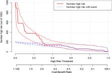
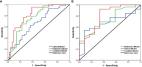
          We downloaded chest CT scans with clear diagnosis of adenocarcinoma and squamous cell carcinoma from The Cancer Imaging Archive (TCIA), extracted 19 imaging features by a radiologist and a thoracic surgeon, including location, spicule, lobulation, cavity, vacuolar sign, necrosis, pleural traction sign, vascular bundle sign, air bronchogram sign, calcification, enhancement degree, distance from pulmonary hilum, atelectasis, pulmonary hilum and bronchial lymph nodes, mediastinal lymph nodes, interlobular septal thickening, pulmonary metastasis, adjacent structures invasion, pleural effusion. Firstly, we apply the glm function of R language to perform logistic univariate analysis on all variables to select variables with P < 0.1. Then, perform logistic multivariate analysis on the selected variables to obtain a predictive model. Next, use the roc function in R language to calculate the AUC value and draw the ROC curve, use the val.prob function in R language to draw the Calibrat curve, and use the rmda package in R language to draw the DCA curve and clinical impact curve. At the same time, 45 patients diagnosed with lung squamous cell carcinoma and lung adenocarcinoma through surgery or biopsy in the Radiotherapy Department and Thoracic Surgery Department of our hospital from 2023 to 2024 were included in the validation group. The chest CT features were jointly determined and recorded by the two doctors mentioned above and included in the validation group. The included image feature data are complete and does not require preprocessing, so directly entering statistical calculations. Perform ROC curves, calibration curves, DCA, and clinical impact curves in the validation group to further validate the predictive model. If the predictive model performs well in the validation group, further draw a nomogram to demonstrate.

          Results

          This study extracted 19 imaging features from the chest CT scans of 75 patients downloaded from TCIA and finally selected 18 complete data for analysis. First, univariate analysis and multivariate analysis were performed, and a total of 5 variables were obtained: spicule, necrosis, air bronchogram Sign, atelectasis, pulmonary hilum and bronchial lymph nodes. After conducting modeling analysis with AUC = 0.887, a validation group was established using clinical cases from our hospital, Draw ROC curve with AUC = 0.865 in the validation group, evaluate the accuracy of the model through Calibrate calibration curve, evaluate the reliability of the model in clinical practice through DCA curve, and further evaluate the practicality of the model in clinical practice through clinical impact curve.

          Conclusion

          It is possible to extract influential features from ordinary chest CT scans to determine lung adenocarcinoma and squamous cell carcinoma. The model we have set up performs well in terms of discrimination, accuracy, reliability, and practicality.

          Related collections

          Most cited references32

          • Record: found
          • Abstract: found
          • Article: not found

          Prognostic value and reproducibility of pretreatment CT texture features in stage III non-small cell lung cancer.

          To determine whether pretreatment CT texture features can improve patient risk stratification beyond conventional prognostic factors (CPFs) in stage III non-small cell lung cancer (NSCLC).
            Bookmark
            • Record: found
            • Abstract: found
            • Article: found
            Is Open Access

            Prediction of pathologic stage in non-small cell lung cancer using machine learning algorithm based on CT image feature analysis

            Purpose To explore imaging biomarkers that can be used for diagnosis and prediction of pathologic stage in non-small cell lung cancer (NSCLC) using multiple machine learning algorithms based on CT image feature analysis. Methods Patients with stage IA to IV NSCLC were included, and the whole dataset was divided into training and testing sets and an external validation set. To tackle imbalanced datasets in NSCLC, we generated a new dataset and achieved equilibrium of class distribution by using SMOTE algorithm. The datasets were randomly split up into a training/testing set. We calculated the importance value of CT image features by means of mean decrease gini impurity generated by random forest algorithm and selected optimal features according to feature importance (mean decrease gini impurity > 0.005). The performance of prediction model in training and testing sets were evaluated from the perspectives of classification accuracy, average precision (AP) score and precision-recall curve. The predictive accuracy of the model was externally validated using lung adenocarcinoma (LUAD) and lung squamous cell carcinoma (LUSC) samples from TCGA database. Results The prediction model that incorporated nine image features exhibited a high classification accuracy, precision and recall scores in the training and testing sets. In the external validation, the predictive accuracy of the model in LUAD outperformed that in LUSC. Conclusions The pathologic stage of patients with NSCLC can be accurately predicted based on CT image features, especially for LUAD. Our findings extend the application of machine learning algorithms in CT image feature prediction for pathologic staging and identify potential imaging biomarkers that can be used for diagnosis of pathologic stage in NSCLC patients. Electronic supplementary material The online version of this article (10.1186/s12885-019-5646-9) contains supplementary material, which is available to authorized users.
              Bookmark
              • Record: found
              • Abstract: found
              • Article: found
              Is Open Access

              Radiomics Study for Predicting the Expression of PD-L1 and Tumor Mutation Burden in Non-Small Cell Lung Cancer Based on CT Images and Clinicopathological Features

              Background The present study compared the predictive performance of pretreatment computed tomography (CT)-based radiomics signatures and clinicopathological and CT morphological factors for ligand programmed death-ligand 1 (PD-L1) expression level and tumor mutation burden (TMB) status and further explored predictive models in patients with advanced-stage non-small cell lung cancer (NSCLC). Methods A total of 120 patients with advanced-stage NSCLC were enrolled in this retrospective study and randomly assigned to a training dataset or validation dataset. Here, 462 radiomics features were extracted from region-of-interest (ROI) segmentation based on pretreatment CT images. The least absolute shrinkage and selection operator (LASSO) and logistic regression were applied to select radiomics features and develop combined models with clinical and morphological factors for PD-L1 expression and TMB status prediction. Ten-fold cross-validation was used to evaluate the accuracy, and the predictive performance of these models was assessed using receiver operating characteristic (ROC) and area under the curve (AUC) analyses. Results The PD-L1-positive expression level correlated with differentiation degree (p = 0.005), tumor shape (p = 0.006), and vascular convergence (p = 0.007). Stage (p = 0.023), differentiation degree (p = 0.017), and vacuole sign (p = 0.016) were associated with TMB status. Radiomics signatures showed good performance for predicting PD-L1 and TMB with AUCs of 0.730 and 0.759, respectively. Predictive models that combined radiomics signatures with clinical and morphological factors dramatically improved the predictive efficacy for PD-L1 (AUC = 0.839) and TMB (p = 0.818). The results were verified in the validation datasets. Conclusions Quantitative CT-based radiomics features have potential value in the classification of PD-L1 expression levels and TMB status. The combined model further improved the predictive performance and provided sufficient information for the guiding of immunotherapy in clinical practice, and it deserves further analysis.
                Bookmark

                Author and article information

                Journal
                Cancer Manag Res
                Cancer Manag Res
                cmar
                Cancer Management and Research
                Dove
                1179-1322
                04 June 2024
                2024
                : 16
                : 547-557
                Affiliations
                [1 ]Department of Radiation Oncology, The Second Hospital of Hebei Medical University , Shijiazhuang, Hebei Province, People’s Republic of China
                [2 ]Department of Thoracic Surgery, The Second Hospital of Hebei Medical University , Shijiazhuang, Hebei Province, People’s Republic of China
                [3 ]Department of Hematology, The Second Hospital of Hebei Medical University , Shijiazhuang, Hebei Province, People’s Republic of China
                Author notes
                Correspondence: Jianmin Luo, Department of Hematology, The Second Hospital of Hebei Medical University , 215 West Heping Road, Shijiazhuang, Hebei Province, People’s Republic of China, Tel +86 1348368527, Email luojm31555@163.com
                Author information
                http://orcid.org/0000-0002-4109-4387
                http://orcid.org/0009-0005-9432-646X
                http://orcid.org/0009-0005-3963-8575
                Article
                462951
                10.2147/CMAR.S462951
                11162187
                38855330
                3cabd84c-55c1-4b1f-825d-db95434e8ea6
                © 2024 Liu et al.

                This work is published and licensed by Dove Medical Press Limited. The full terms of this license are available at https://www.dovepress.com/terms.php and incorporate the Creative Commons Attribution – Non Commercial (unported, v3.0) License ( http://creativecommons.org/licenses/by-nc/3.0/). By accessing the work you hereby accept the Terms. Non-commercial uses of the work are permitted without any further permission from Dove Medical Press Limited, provided the work is properly attributed. For permission for commercial use of this work, please see paragraphs 4.2 and 5 of our Terms ( https://www.dovepress.com/terms.php).

                History
                : 05 February 2024
                : 25 May 2024
                Page count
                Figures: 8, Tables: 4, References: 33, Pages: 11
                Funding
                Funded by: This work was supported by the Health of Hebei Province Commission for medical scientific research funding of China;
                This work was supported by the Health of Hebei Province Commission for medical scientific research funding of China (No. 20180396).
                Categories
                Original Research

                Oncology & Radiotherapy
                lung cancer,luad,lscc,image features,predict
                Oncology & Radiotherapy
                lung cancer, luad, lscc, image features, predict

                Comments

                Comment on this article