704
views
0
recommends
+1 Recommend
0 collections
    3
    shares
      • Record: found
      • Abstract: found
      • Article: found
      Is Open Access

      FIP200, a ULK-interacting protein, is required for autophagosome formation in mammalian cells

      research-article

      Read this article at

      Bookmark
          There is no author summary for this article yet. Authors can add summaries to their articles on ScienceOpen to make them more accessible to a non-specialist audience.

          Abstract

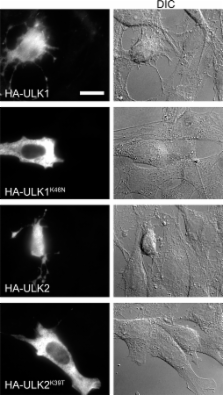
          Autophagy is a membrane-mediated intracellular degradation system. The serine/threonine kinase Atg1 plays an essential role in autophagosome formation. However, the role of the mammalian Atg1 homologues UNC-51–like kinase (ULK) 1 and 2 are not yet well understood. We found that murine ULK1 and 2 localized to autophagic isolation membrane under starvation conditions. Kinase-dead alleles of ULK1 and 2 exerted a dominant-negative effect on autophagosome formation, suggesting that ULK kinase activity is important for autophagy. We next screened for ULK binding proteins and identified the focal adhesion kinase family interacting protein of 200 kD (FIP200), which regulates diverse cellular functions such as cell size, proliferation, and migration. We found that FIP200 was redistributed from the cytoplasm to the isolation membrane under starvation conditions. In FIP200-deficient cells, autophagy induction by various treatments was abolished, and both stability and phosphorylation of ULK1 were impaired. These results suggest that FIP200 is a novel mammalian autophagy factor that functions together with ULKs.

          Related collections

          Most cited references90

          • Record: found
          • Abstract: found
          • Article: not found

          RADIOAUTOGRAPHIC STUDIES OF CHOLINE INCORPORATION INTO PERIPHERAL NERVE MYELIN

          This radioautographic study was designed to localize the cytological sites involved in the incorporation of a lipid precursor into the myelin and the myelin-related cell of the peripheral nervous system. Both myelinating and fully myelinated cultures of rat dorsal root ganglia were exposed to a 30-min pulse of tritiated choline and either fixed immediately or allowed 6 or 48 hr of chase incubation before fixation. After Epon embedding, light and electron microscopic radioautograms were prepared with Ilford L-4 emulsion. Analysis of the pattern of choline incorporation into myelinating cultures indicated that radioactivity appeared all along the length of the internode, without there being a preferential site of initial incorporation. Light microscopic radioautograms of cultures at varying states of maturity were compared in order to determine the relative degree of myelin labeling. This analysis indicated that the myelin-Schwann cell unit in the fully myelinated cultures incorporated choline as actively as did this unit in the myelinating cultures. Because of technical difficulties, it was not possible to determine the precise localization of the incorporated radioactivity within the compact myelin. These data are related to recent biochemical studies indicating that the mature myelin of the central nervous system does incorporate a significant amount of lipid precursor under the appropriate experimental conditions. These observations support the concept that a significant amount of myelin-related metabolic activity occurs in mature tissue; this activity is considered part of an essential and continuous process of myelin maintenance and repair.
            Bookmark
            • Record: found
            • Abstract: found
            • Article: not found

            Systematic identification of protein complexes in Saccharomyces cerevisiae by mass spectrometry.

            The recent abundance of genome sequence data has brought an urgent need for systematic proteomics to decipher the encoded protein networks that dictate cellular function. To date, generation of large-scale protein-protein interaction maps has relied on the yeast two-hybrid system, which detects binary interactions through activation of reporter gene expression. With the advent of ultrasensitive mass spectrometric protein identification methods, it is feasible to identify directly protein complexes on a proteome-wide scale. Here we report, using the budding yeast Saccharomyces cerevisiae as a test case, an example of this approach, which we term high-throughput mass spectrometric protein complex identification (HMS-PCI). Beginning with 10% of predicted yeast proteins as baits, we detected 3,617 associated proteins covering 25% of the yeast proteome. Numerous protein complexes were identified, including many new interactions in various signalling pathways and in the DNA damage response. Comparison of the HMS-PCI data set with interactions reported in the literature revealed an average threefold higher success rate in detection of known complexes compared with large-scale two-hybrid studies. Given the high degree of connectivity observed in this study, even partial HMS-PCI coverage of complex proteomes, including that of humans, should allow comprehensive identification of cellular networks.
              Bookmark
              • Record: found
              • Abstract: found
              • Article: not found

              How to interpret LC3 immunoblotting.

              Microtubule-associated protein light chain 3 (LC3) is now widely used to monitor autophagy. One approach is to detect LC3 conversion (LC3-I to LC3-II) by immunoblot analysis because the amount of LC3-II is clearly correlated with the number of autophagosomes. However, LC3-II itself is degraded by autophagy, making interpretation of the results of LC3 immunoblotting problematic. Furthermore, the amount of LC3 at a certain time point does not indicate autophagic flux, and therefore, it is important to measure the amount of LC3-II delivered to lysosomes by comparing LC3-II levels in the presence and absence of lysosomal protease inhibitors. Another problem with this method is that LC3-II tends to be much more sensitive to be detected by immunoblotting than LC3-I. Accordingly, simple comparison of LC3-I and LC3-II, or summation of LC3-I and LC3-II for ratio determinations, may not be appropriate, and rather, the amount of LC3-II can be compared between samples.
                Bookmark

                Author and article information

                Journal
                J Cell Biol
                jcb
                The Journal of Cell Biology
                The Rockefeller University Press
                0021-9525
                1540-8140
                5 May 2008
                : 181
                : 3
                : 497-510
                Affiliations
                [1 ]Department of Physiology and Cell Biology, Tokyo Medical and Dental University, Bunkyo-ku, Tokyo 113-8549, Japan
                [2 ]Biological Information Research Center, National Institutes of Advanced Industrial Science and Technology, Kohtoh-ku, Tokyo 135-0064, Japan
                [3 ]Department of Internal Medicine-MMG, University of Michigan Medical School, Ann Arbor, MI 48109
                [4 ]Solution Oriented Research for Science and Technology, Japan Science and Technology Agency, Kawaguchi, Saitama 332-0012, Japan
                Author notes

                Correspondence to Noboru Mizushima: nmizu.phy2@ 123456tmd.ac.jp

                Article
                200712064
                10.1083/jcb.200712064
                2364687
                18443221
                3c0cdceb-8232-408d-a4d1-aa7bc7c61c52
                © 2008 Hara et al.

                This article is distributed under the terms of an Attribution–Noncommercial–Share Alike–No Mirror Sites license for the first six months after the publication date (see http://www.jcb.org/misc/terms.shtml). After six months it is available under a Creative Commons License (Attribution–Noncommercial–Share Alike 3.0 Unported license, as described at http://creativecommons.org/licenses/by-nc-sa/3.0/).

                History
                : 12 December 2007
                : 2 April 2008
                Categories
                Research Articles
                Article

                Cell biology
                Cell biology

                Comments

                Comment on this article