23
views
0
recommends
+1 Recommend
0 collections
    0
    shares
      • Record: found
      • Abstract: found
      • Article: found
      Is Open Access

      Definitive 3D-CRT for clinically localized prostate cancer: modifications of the clinical target volume following a prostate MRI and the clinical benefits

      research-article

      Read this article at

      Bookmark
          There is no author summary for this article yet. Authors can add summaries to their articles on ScienceOpen to make them more accessible to a non-specialist audience.

          Abstract

          Purpose

          To evaluate the modifications of the tumor stage and clinical target volume following a prostate magnetic resonance imaging (MRI) to evaluate the tumor (T) staging, and the clinical benefits for prostate cancer.

          Methods

          A total of 410 patients with newly diagnosed and clinically localized prostate cancer were retrospectively analyzed. The patients were treated with definitive three-dimensional conformal radiotherapy (3D-CRT). In all of the patients, digital rectal examination, transrectal ultrasound, prostate biopsy and computed tomography were performed to evaluate the clinical stage. Of the 410 patients, 189 patients had undergone a prostate MRI study to evaluate the T staging, and 221 patients had not.

          Results

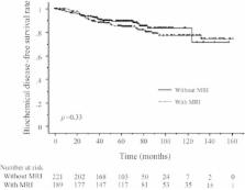
          Modification of the T stage after the prostate MRI was seen in 39 (25%) of the 157 evaluable patients, and a modification of the risk group was made in 14 (9%) patients. Eventually, a modification of the CTV in 3D-CRT planning was made in 13 (8%) patients, and 10 of these had extracapsular disease. Most of the other modifications of the T staging were associated with intracapsular lesions of prostate cancer which did not change the CTV. There were no significant differences in the biological relapse-free survival between the patients with and without a prostate MRI study.

          Conclusions

          Modification of the CTV were recognized in only 8% of the patients, most of whom had extracapsular disease, although that of the T stage was seen in approximately one-quarter of the patients. Prostate MRI should only be selected for patients with a high probability of extracapsular involvement.

          Related collections

          Most cited references28

          • Record: found
          • Abstract: found
          • Article: not found

          Defining biochemical failure following radiotherapy with or without hormonal therapy in men with clinically localized prostate cancer: recommendations of the RTOG-ASTRO Phoenix Consensus Conference.

          In 1996 the American Society for Therapeutic Radiology and Oncology (ASTRO) sponsored a Consensus Conference to establish a definition of biochemical failure after external beam radiotherapy (EBRT). The ASTRO definition defined prostate specific antigen (PSA) failure as occurring after three consecutive PSA rises after a nadir with the date of failure as the point halfway between the nadir date and the first rise or any rise great enough to provoke initiation of therapy. This definition was not linked to clinical progression or survival; it performed poorly in patients undergoing hormonal therapy (HT), and backdating biased the Kaplan-Meier estimates of event-free survival. A second Consensus Conference was sponsored by ASTRO and the Radiation Therapy Oncology Group in Phoenix, Arizona, on January 21, 2005, to revise the ASTRO definition. The panel recommended: (1) a rise by 2 ng/mL or more above the nadir PSA be considered the standard definition for biochemical failure after EBRT with or without HT; (2) the date of failure be determined "at call" (not backdated). They recommended that investigators be allowed to use the ASTRO Consensus Definition after EBRT alone (no hormonal therapy) with strict adherence to guidelines as to "adequate follow-up." To avoid the artifacts resulting from short follow-up, the reported date of control should be listed as 2 years short of the median follow-up. For example, if the median follow-up is 5 years, control rates at 3 years should be cited. Retaining a strict version of the ASTRO definition would allow comparisons with a large existing body of literature.
            Bookmark
            • Record: found
            • Abstract: found
            • Article: not found

            Advances in magnetic resonance imaging: how they are changing the management of prostate cancer.

            Although magnetic resonance imaging (MRI) is emerging as the most commonly used imaging modality for prostate cancer (PCa) detection, treatment planning, and follow-up, its acceptance has not been uniform. Recently, great interest has been shown in multiparametric MRI, which combines anatomic T2-weighted (T2W) imaging with MR spectroscopic imaging (MRSI), dynamic contrast-enhanced MRI (DCE-MRI), and diffusion-weighted imaging (DWI). The aim of this article is to review the current roles of these MR techniques in different aspects of PCa management: initial diagnosis, biopsy strategies, planning of radical prostatectomy (RP) and external radiation therapy (RT), and implementation of alternative focal therapies. The authors searched the Medline and Cochrane Library databases (primary fields: prostatic neoplasm, magnetic resonance). The search was performed without language restriction from January 2008 to November 2010. Initial diagnosis: The data suggest that the combination of T2W MRI and DWI or MRSI with DCE-MRI has the potential to guide biopsy to the most aggressive cancer foci in patients with previously negative biopsies, increasing the accuracy of the procedure. Transrectal MR-guided prostate biopsy can improve PCa detection, but its availability is still limited and the examination time is rather long. Planning of RP: It appears that adding MRSI, DWI, and/or DCE-MRI to T2W MRI can facilitate better preoperative characterization of cancer with regard to location, size, and relationship to prostatic and extraprostatic structures, and it may also facilitate early detection of local recurrence. Thus, use of these MR techniques may improve surgical, oncologic, and functional management. Planning of external RT and focal therapies: MR techniques have similar potential in these areas, but the published data remain very limited. MRI technology is continuously evolving, and more extensive use of MRI technology in clinical trials and practice will help to improve PCa diagnosis and treatment planning. Copyright © 2011 European Association of Urology. Published by Elsevier B.V. All rights reserved.
              Bookmark
              • Record: found
              • Abstract: found
              • Article: not found

              Prostate cancer: body-array versus endorectal coil MR imaging at 3 T--comparison of image quality, localization, and staging performance.

              To prospectively compare image quality and accuracy of prostate cancer localization and staging with body-array coil (BAC) versus endorectal coil (ERC) T2-weighted magnetic resonance (MR) imaging at 3 T, with histopathologic findings as the reference standard. After institutional review board approval and written informed consent, 46 men underwent 3-T T2-weighted MR imaging with a BAC (voxel size, 0.43 x 0.43 x 4.00 mm) and an ERC (voxel size, 0.26 x 0.26 x 2.50 mm) before radical prostatectomy. Four radiologists independently evaluated data sets obtained with the BAC and ERC separately. Ten image quality characteristics related to prostate cancer localization and staging were assigned scores. Prostate cancer presence was recorded with a five-point probability scale in each of 14 segments that included the whole prostate. Disease stage was classified as organ-confined or locally advanced with a five-point probability scale. Whole-mount-section histopathologic examination was the reference standard. Areas under the receiver operating characteristic curve (AUCs) and diagnostic performance parameters were determined. A difference with a P value of less than .05 was considered significant. Forty-six patients (mean age, 61 years) were included for analysis. Significantly more motion artifacts were present with ERC imaging (P<.001). All other image quality characteristics improved significantly (P<.001) with ERC imaging. With ERC imaging, the AUC for localization of prostate cancer was significantly increased from 0.62 to 0.68 (P<.001). ERC imaging significantly increased the AUCs for staging, and sensitivity for detection of locally advanced disease by experienced readers was increased from 7% (one of 15) to a range of 73% (11 of 15) to 80% (12 of 15) (P<.05), whereas a high specificity of 97% (30 of 31) to 100% (31 of 31) was maintained. Extracapsular extension as small as 0.5 mm at histopathologic examination could be accurately detected only with ERC imaging. Image quality and localization improved significantly with ERC imaging compared with BAC imaging. For experienced radiologists, the staging performance was significantly better with ERC imaging. (c) RSNA, 2007.
                Bookmark

                Author and article information

                Contributors
                specialvirtue38@yahoo.co.jp
                ogurieye@med.uoeh-u.ac.jp
                fujii-m@med.uoeh-u.ac.jp
                k-yahara@med.uoeh-u.ac.jp
                yhlinda@med.uoeh-u.ac.jp
                n-fuji@med.uoeh-u.ac.jp
                ykorogi@med.uoeh-u.ac.jp
                Journal
                Springerplus
                Springerplus
                SpringerPlus
                Springer International Publishing (Cham )
                2193-1801
                15 July 2015
                15 July 2015
                2015
                : 4
                : 347
                Affiliations
                [ ]Department of Radiology, Kitakyushu General Hospital, Kitakyushu, Japan
                [ ]Department of Radiology, University of Occupational and Environmental Health, 1-1 Iseigaoka, Yahatanishi-ku, Kitakyushu, 807-8555 Japan
                [ ]Department of Urology, University of Occupational and Environmental Health, Kitakyushu, Japan
                Article
                1138
                10.1186/s40064-015-1138-9
                4502053
                3bf9d6f8-59c1-4635-a76b-8a43f1d16b5c
                © Yamaguchi et al. 2015

                Open AccessThis article is distributed under the terms of the Creative Commons Attribution 4.0 International License ( http://creativecommons.org/licenses/by/4.0/), which permits unrestricted use, distribution, and reproduction in any medium, provided you give appropriate credit to the original author(s) and the source, provide a link to the Creative Commons license, and indicate if changes were made.

                History
                : 9 March 2015
                : 6 July 2015
                Categories
                Research
                Custom metadata
                © The Author(s) 2015

                Uncategorized
                prostate cancer,magnetic resonance imaging,clinical target volume,tumor staging,three-dimensional conformal radiotherapy

                Comments

                Comment on this article