3
views
0
recommends
+1 Recommend
0 collections
    0
    shares
      • Record: found
      • Abstract: found
      • Article: found
      Is Open Access

      Comparative Evaluation of Four Different Obturating Techniques in Primary Teeth Using Cone-beam Computed Tomography: An In Vivo Study

      research-article

      Read this article at

      Bookmark
          There is no author summary for this article yet. Authors can add summaries to their articles on ScienceOpen to make them more accessible to a non-specialist audience.

          A bstract

          Background

          The quality of the obturation plays a significant role in the success of endodontic treatment. To date, various technologies have been used to evaluate the quality of obturation, but all of them have their own limitations. In order to overcome those limitations, recent technological advancements like cone-beam computed tomography (CBCT) can be helpful.

          Aim

          To compare and evaluate the efficiency of different root canal obturation techniques in primary teeth using CBCT.

          Materials and methods

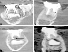
          A total of 80 root canals in 30 children aged between 4 and 9 years were selected and divided into four groups, with 20 root canals in each. Obturation in group I was performed using the endodontic pressure syringe; group II—hand spreaders; group III—Lentulo spirals mounted on slow-speed handpiece; and group IV—insulin syringe. The quality of obturation was evaluated using a CBCT scan.

          Results

          Group I samples showed the most optimally filled canals followed by II and III; least in group IV. A maximum number of overfilled canals was exhibited in group III samples. Voids were minimal in all four groups and the values obtained were not statistically significant.

          Conclusion

          Obturation with an endodontic pressure syringe reported the highest number of optimally filled root canals and the insulin syringe showed the least number of optimally filled canals.

          How to cite this article

          Ali SM, Mukthineni S, Sai Sankar AJ, et al. Comparative Evaluation of Four Different obturating Techniques in Primary Teeth using Cone-beam Computed Tomography: An In Vivo Study. Int J Clin Pediatr Dent 2023;16(2):218-222.

          Related collections

          Most cited references29

          • Record: found
          • Abstract: found
          • Article: not found

          Predicting pulpectomy success and its relationship to exfoliation and succedaneous dentition.

          This study evaluated factors that affected pulpectomy (PE) success and its effect on the succedaneous tooth's eruption and enamel formation. Sixty-five of 250 patients with PEs met the selection criteria and yielded 81 zinc oxide-eugenol PEs (30 incisors, 51 molars) followed a mean time of 90.8 months. Overall PE success was 77.7% with no difference between molars and incisors (P = 0.53). Enamel defects were observed in 18.7% of succedaneous teeth and were related (P = 0.005) to the pre-existing infection causing excess root resorption (>1 mm preoperative root resorption = 44.4% defects) but were not related to overretention of ZOE filler (P = 1) or length of fill (P = 0.36). The PE procedure was not related to causing succedaneous tooth defects since teeth replacing PEs showed no significant increase in the incidence of defects compared with untreated contralateral controls (P = 0.99). There was a 20% incidence of succedaneous tooth anterior cross-bite or palatal eruption following incisor PEs and 21.6% ectopic eruption of premolars following primary molar PEs. Most PEs (95.9%) were lost at their normal exfoliation time or earlier, but 35.8% needed extraction due to overretention by soft tissue at the time of shedding. Pulpectomy success rates showed that the most important preoperative predictor was the amount of primary tooth root resorption. Greater than 1 mm of root resorption resulted in only a 23.1% success rate, which was significant (P = 0.001). Pulpectomies filled short or to the apex had a significantly greater success (P = 0.011) than long fills. Pulpectomies correctly done do not appear to contribute to adverse effects on succedaneous tooth formation but have a 20% chance of altering the path of permanent tooth eruption.
            Bookmark
            • Record: found
            • Abstract: found
            • Article: found
            Is Open Access

            Cone Beam Computed Tomography - Know its Secrets

            Cone-beam computed tomography (CBCT) is an advanced imaging modality that has high clinical applications in the field of dentistry. CBCT proved to be a successful investigative modality that has been used for dental and maxillofacial imaging. Radiation exposure dose from CBCT is 10 times less than from conventional CT scans during maxillofacial exposure. Furthermore, CBCT is highly accurate and can provide a three-dimensional volumetric data in axial, sagittal and coronal planes. This article describes the basic technique, difference in CBCT from CT and main clinical applications of CBCT.
              Bookmark
              • Record: found
              • Abstract: found
              • Article: not found

              Comparison of different obturation techniques for primary molars by digital radiography.

              The purpose of this study was to compare six methods of root canal filling in primary mandibular second molars via digital radiography.
                Bookmark

                Author and article information

                Journal
                Int J Clin Pediatr Dent
                Int J Clin Pediatr Dent
                IJCPD
                International Journal of Clinical Pediatric Dentistry
                Jaypee Brothers Medical Publishers
                0974-7052
                0975-1904
                Mar-Apr 2023
                : 16
                : 2
                : 218-222
                Affiliations
                [1-5 ]Department of Pediatric Dentistry, Sibar Institute of Dental Sciences (SIDS), Guntur, Andhra Pradesh, India
                [6 ]Department of Oral Medicine and Radiology, Sibar Institute of Dental Sciences (SIDS), Guntur, Andhra Pradesh, India
                Author notes
                AJ Sai Sankar, Department of Pediatric Dentistry, Sibar Institute of Dental Sciences (SIDS), Guntur, Andhra Pradesh, India, Phone: +91 9346550646, e-mail: saisamata@ 123456gmail.com
                Author information
                https://orcid.org/0000-0001-9363-4317
                https://orcid.org/0000-0002-6233-6963
                https://orcid.org/0000-0003-4720-6404
                https://orcid.org/0000-0001-7572-9873
                https://orcid.org/0000-0003-1603-2253
                Article
                10.5005/jp-journals-10005-2566
                10373754
                3be659f2-58c1-4d96-844d-7150681f073c
                Copyright © 2023; The Author(s).

                © The Author(s). 2023 Open Access This article is distributed under the terms of the Creative Commons Attribution 4.0 International License ( https://creativecommons.org/licenses/by-nc/4.0/), which permits unrestricted use, distribution, and non-commercial reproduction in any medium, provided you give appropriate credit to the original author(s) and the source, provide a link to the Creative Commons license, and indicate if changes were made. The Creative Commons Public Domain Dedication waiver ( http://creativecommons.org/publicdomain/zero/1.0/) applies to the data made available in this article, unless otherwise stated.

                History
                Categories
                Original Research

                cone-beam computed tomography,obturation,pulpectomy,zinc oxide eugenol

                Comments

                Comment on this article