0
views
0
recommends
+1 Recommend
0 collections
    0
    shares
      • Record: found
      • Abstract: found
      • Article: found
      Is Open Access

      Cortical Networks Relating to Arousal Are Differentially Coupled to Neural Activity and Hemodynamics

      research-article

      Read this article at

      Bookmark
          There is no author summary for this article yet. Authors can add summaries to their articles on ScienceOpen to make them more accessible to a non-specialist audience.

          Abstract

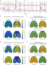
          Even in the absence of specific sensory input or a behavioral task, the brain produces structured patterns of activity. This organized activity is modulated by changes in arousal. Here, we use wide-field voltage imaging to establish how arousal relates to cortical network voltage and hemodynamic activity in spontaneously behaving head-fixed male and female mice expressing the voltage-sensitive fluorescent FRET sensor Butterfly 1.2. We find that global voltage and hemodynamic signals are both positively correlated with changes in arousal with a maximum correlation of 0.5 and 0.25, respectively, at a time lag of 0 s. We next show that arousal influences distinct cortical regions for both voltage and hemodynamic signals. These include a broad positive correlation across most sensory-motor cortices extending posteriorly to the primary visual cortex observed in both signals. In contrast, activity in the prefrontal cortex is positively correlated to changes in arousal for the voltage signal while it is a slight net negative correlation observed in the hemodynamic signal. Additionally, we show that coherence between voltage and hemodynamic signals relative to arousal is strongest for slow frequencies below 0.15 Hz and is near zero for frequencies >1 Hz. We finally show that coupling patterns are dependent on the behavioral state of the animal with correlations being driven by periods of increased orofacial movement. Our results indicate that while hemodynamic signals show strong relations to behavior and arousal, these relations are distinct from those observed by voltage activity.

          Related collections

          Most cited references49

          • Record: found
          • Abstract: found
          • Article: not found

          The brain's default network: anatomy, function, and relevance to disease.

          Thirty years of brain imaging research has converged to define the brain's default network-a novel and only recently appreciated brain system that participates in internal modes of cognition. Here we synthesize past observations to provide strong evidence that the default network is a specific, anatomically defined brain system preferentially active when individuals are not focused on the external environment. Analysis of connectional anatomy in the monkey supports the presence of an interconnected brain system. Providing insight into function, the default network is active when individuals are engaged in internally focused tasks including autobiographical memory retrieval, envisioning the future, and conceiving the perspectives of others. Probing the functional anatomy of the network in detail reveals that it is best understood as multiple interacting subsystems. The medial temporal lobe subsystem provides information from prior experiences in the form of memories and associations that are the building blocks of mental simulation. The medial prefrontal subsystem facilitates the flexible use of this information during the construction of self-relevant mental simulations. These two subsystems converge on important nodes of integration including the posterior cingulate cortex. The implications of these functional and anatomical observations are discussed in relation to possible adaptive roles of the default network for using past experiences to plan for the future, navigate social interactions, and maximize the utility of moments when we are not otherwise engaged by the external world. We conclude by discussing the relevance of the default network for understanding mental disorders including autism, schizophrenia, and Alzheimer's disease.
            Bookmark
            • Record: found
            • Abstract: found
            • Article: not found

            Functional connectivity in the motor cortex of resting human brain using echo-planar MRI.

            An MRI time course of 512 echo-planar images (EPI) in resting human brain obtained every 250 ms reveals fluctuations in signal intensity in each pixel that have a physiologic origin. Regions of the sensorimotor cortex that were activated secondary to hand movement were identified using functional MRI methodology (FMRI). Time courses of low frequency (< 0.1 Hz) fluctuations in resting brain were observed to have a high degree of temporal correlation (P < 10(-3)) within these regions and also with time courses in several other regions that can be associated with motor function. It is concluded that correlation of low frequency fluctuations, which may arise from fluctuations in blood oxygenation or flow, is a manifestation of functional connectivity of the brain.
              Bookmark
              • Record: found
              • Abstract: found
              • Article: not found

              Spontaneous behaviors drive multidimensional, brainwide activity

              Neuronal populations in sensory cortex produce variable responses to sensory stimuli and exhibit intricate spontaneous activity even without external sensory input. Cortical variability and spontaneous activity have been variously proposed to represent random noise, recall of prior experience, or encoding of ongoing behavioral and cognitive variables. Recording more than 10,000 neurons in mouse visual cortex, we observed that spontaneous activity reliably encoded a high-dimensional latent state, which was partially related to the mouse’s ongoing behavior and was represented not just in visual cortex but also across the forebrain. Sensory inputs did not interrupt this ongoing signal but added onto it a representation of external stimuli in orthogonal dimensions. Thus, visual cortical population activity, despite its apparently noisy structure, reliably encodes an orthogonal fusion of sensory and multidimensional behavioral information.
                Bookmark

                Author and article information

                Journal
                J Neurosci
                J Neurosci
                jneuro
                J. Neurosci
                The Journal of Neuroscience
                Society for Neuroscience
                0270-6474
                1529-2401
                20 May 2024
                19 June 2024
                19 June 2024
                : 44
                : 25
                : e0298232024
                Affiliations
                [1] 1 Department of Biology, Emory University, Atlanta, Georgia 30322
                [2] 2Department of Biomedical Engineering, Emory and Georgia Tech , Atlanta, Georgia 30322
                Author notes

                Author contributions: A.E.M., L.M-B., and D.J. designed research; A.E.M., Y.W., and B.L.C. performed research; L.M.-B., A.E.M., Y.W., S.D.K., and D.J. analyzed data; L.M.-B., A.E.M., P.Y.B., S.D.K., G.B.S., and D.J. wrote the paper.

                We thank Dr. Thomas Knöpfel for sharing the VSFP 1.2 construct along with valuable advice on their use. We thank Dr. Hongkui Zeng for providing the initial VSFP mice for our colony. This work was supported by National Institute of Neurological Disorders and Stroke (UO1NS094302, RO1NS078095, and 1S10OD016244), National Institutes of Health BRAIN Initiative (R01 NS111470), National Institute of Biomedical Imaging and Bioengineering (T32EB025816 and R01eb029857), and Eunice Kennedy Shriver National Institute of Child Health and Human Development (NICHD; T32HD071845).

                The authors declare no competing financial interests.

                Correspondence should be addressed to Dieter Jaeger at djaeger@ 123456emory.edu .
                Author information
                https://orcid.org/0000-0002-1856-7713
                https://orcid.org/0000-0002-7523-1247
                https://orcid.org/0000-0001-8194-4166
                https://orcid.org/0000-0001-5737-1660
                https://orcid.org/0000-0003-2039-7706
                https://orcid.org/0000-0002-5122-1319
                Article
                jneuro-44-e0298232024
                10.1523/JNEUROSCI.0298-23.2024
                11209646
                38769007
                3bc0f5da-5d7d-4170-8212-148a2dd41def
                Copyright © 2024 Meyer-Baese et al.

                This is an open-access article distributed under the terms of the Creative Commons Attribution 4.0 International license, which permits unrestricted use, distribution and reproduction in any medium provided that the original work is properly attributed.

                History
                : 17 February 2023
                : 6 May 2024
                : 9 May 2024
                Funding
                Funded by: HHS | NIH | National Institute of Neurological Disorders and Stroke (NINDS)
                Award ID: UO1NS094302
                Award ID: R01NS078095
                Award ID: R01NS111470
                Funded by: HHS | NIH | National Institute of Biomedical Imaging and Bioengineering (NIBIB)
                Award ID: R01EB029857
                Award ID: T32EB025816
                Funded by: HHS | NIH | Eunice Kennedy Shriver National Institute of Child Health and Human Development (NICHD)
                Award ID: T32HD071845
                Categories
                Research Articles
                Systems/Circuits

                cortex,fmri,mouse,pupil diameter,voltage imaging,wide-field optical imaging

                Comments

                Comment on this article