26
views
0
recommends
+1 Recommend
0 collections
    0
    shares
      • Record: found
      • Abstract: found
      • Article: found
      Is Open Access

      Cancer-Associated Fibroblasts from Hepatocellular Carcinoma Promote Malignant Cell Proliferation by HGF Secretion

      research-article

      Read this article at

      Bookmark
          There is no author summary for this article yet. Authors can add summaries to their articles on ScienceOpen to make them more accessible to a non-specialist audience.

          Abstract

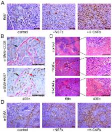
          Cancer-associated fibroblasts (CAFs) are reported to support tumorigenesis by stimulating angiogenesis, cancer cell proliferation, and invasion in most solid tumors. However, the roles of CAFs in the liver cancer microenvironment have not been thoroughly studied. In our previous study, we successfully isolated CAFs from hepatocellular carcinoma (HCC) (H-CAFs) and proved that H-CAFs suppressed the activation of NK cells and thereby created favorable conditions for HCC progression. In our present study, we found that the proliferation of MHCC97L and Hep3B cells was significantly promoted by treatment with conditioned medium from H-CAFs. Pathological analysis also revealed that H-CAFs increased the proportion of Ki-67 (+) malignant cells and prevented them from undergoing necrosis. Moreover, the concentration of hepatocyte growth factor (HGF) cytokine in the conditioned medium of H-CAFs was higher than conditioned medium from normal skin fibroblasts (NSFs). Anti-HGF significantly reduced the proliferation-promoting capability of H-CAFs. In addition, we found that the abundance of H-CAFs correlated positively with tumor size. These results indicate that H-CAFs are an important factor for promoting the growth of HCC in vitro and in vivo, and that HGF plays a key role in HCC proliferation induced by H-CAFs.

          Related collections

          Most cited references20

          • Record: found
          • Abstract: found
          • Article: not found

          Cancer-associated stromal fibroblasts promote pancreatic tumor progression.

          Pancreatic adenocarcinoma is characterized by a dense background of tumor associated stroma originating from abundant pancreatic stellate cells. The aim of this study was to determine the effect of human pancreatic stellate cells (HPSC) on pancreatic tumor progression. HPSCs were isolated from resected pancreatic adenocarcinoma samples and immortalized with telomerase and SV40 large T antigen. Effects of HPSC conditioned medium (HPSC-CM) on in vitro proliferation, migration, invasion, soft-agar colony formation, and survival in the presence of gemcitabine or radiation therapy were measured in two pancreatic cancer cell lines. The effects of HPSCs on tumors were examined in an orthotopic murine model of pancreatic cancer by co-injecting them with cancer cells and analyzing growth and metastasis. HPSC-CM dose-dependently increased BxPC3 and Panc1 tumor cell proliferation, migration, invasion, and colony formation. Furthermore, gemcitabine and radiation therapy were less effective in tumor cells treated with HPSC-CM. HPSC-CM activated the mitogen-activated protein kinase and Akt pathways in tumor cells. Co-injection of tumor cells with HPSCs in an orthotopic model resulted in increased primary tumor incidence, size, and metastasis, which corresponded with the proportion of HPSCs. HPSCs produce soluble factors that stimulate signaling pathways related to proliferation and survival of pancreatic cancer cells, and the presence of HPSCs in tumors increases the growth and metastasis of these cells. These data indicate that stellate cells have an important role in supporting and promoting pancreatic cancer. Identification of HPSC-derived factors may lead to novel stroma-targeted therapies for pancreatic cancer.
            Bookmark
            • Record: found
            • Abstract: found
            • Article: found
            Is Open Access

            Cancer Associated Fibroblasts Promote Tumor Growth and Metastasis by Modulating the Tumor Immune Microenvironment in a 4T1 Murine Breast Cancer Model

            Background Local inflammation associated with solid tumors commonly results from factors released by tumor cells and the tumor stroma, and promotes tumor progression. Cancer associated fibroblasts comprise a majority of the cells found in tumor stroma and are appealing targets for cancer therapy. Here, our aim was to determine the efficacy of targeting cancer associated fibroblasts for the treatment of metastatic breast cancer. Methodology/Principal Findings We demonstrate that cancer associated fibroblasts are key modulators of immune polarization in the tumor microenvironment of a 4T1 murine model of metastatic breast cancer. Elimination of cancer associated fibroblasts in vivo by a DNA vaccine targeted to fibroblast activation protein results in a shift of the immune microenvironment from a Th2 to Th1 polarization. This shift is characterized by increased protein expression of IL-2 and IL-7, suppressed recruitment of tumor-associated macrophages, myeloid derived suppressor cells, T regulatory cells, and decreased tumor angiogenesis and lymphangiogenesis. Additionally, the vaccine improved anti-metastatic effects of doxorubicin chemotherapy and enhanced suppression of IL-6 and IL-4 protein expression while increasing recruitment of dendritic cells and CD8+ T cells. Treatment with the combination therapy also reduced tumor-associated Vegf, Pdgfc, and GM-CSF mRNA and protein expression. Conclusions/Significance Our findings demonstrate that cancer associated fibroblasts promote tumor growth and metastasis through their role as key modulators of immune polarization in the tumor microenvironment and are valid targets for therapy of metastatic breast cancer.
              Bookmark
              • Record: found
              • Abstract: found
              • Article: not found

              Comparison of metabolic pathways between cancer cells and stromal cells in colorectal carcinomas: a metabolic survival role for tumor-associated stroma.

              Understanding tumor metabolism is important for the development of anticancer therapies. Immunohistochemical evaluation of colorectal adenocarcinomas showed that cancer cells share common enzyme/transporter activities suggestive of an anaerobic metabolism [high lactate dehydrogenase 5 (LDH5)/hypoxia-inducible factor alphas (HIFalphas)] with high ability for glucose absorption and lactate extrusion [high glucose transporter 1 (GLUT1)/monocarboxylate transporter (MCT1)]. The tumor-associated fibroblasts expressed proteins involved in lactate absorption (high MCT1/MCT2), lactate oxidation (high LDH1 and low HIFalphas/LDH5), and reduced glucose absorption (low GLUT1). The expression profile of the tumor-associated endothelium indicated aerobic metabolism (high LDH1 and low HIFalphas/LDH5), high glucose absorption (high GLUT1), and resistance to lactate intake (lack of MCT1). It is suggested that the newly formed stroma and vasculature express complementary metabolic pathways, buffering and recycling products of anaerobic metabolism to sustain cancer cell survival. Tumors survive and grow because they are capable of organizing the regional fibroblasts and endothelial cells into a harmoniously collaborating metabolic domain.
                Bookmark

                Author and article information

                Contributors
                Role: Editor
                Journal
                PLoS One
                PLoS ONE
                plos
                plosone
                PLoS ONE
                Public Library of Science (San Francisco, USA )
                1932-6203
                2013
                7 May 2013
                : 8
                : 5
                : e63243
                Affiliations
                [1 ]Department of Hepatic Surgery, The Third Affiliated Hospital of Sun Yat-sen University, Guangzhou, China
                [2 ]Guangdong Provincial Key Laboratory of Liver Disease Research, Guangzhou, China
                [3 ]Department of Medical Oncology, The Third Affiliated Hospital of Sun Yat-sen University, Guangzhou, China
                [4 ]Cell-gene Therapy Translational Medicine Research Center, The Third Affiliated Hospital of Sun Yat-sen University, Guangzhou, China
                [5 ]Department of Hepatobiliary Surgery, Nanfang Hospital, Southern Medical University, Guangzhou, China
                University of Southern California, United States of America
                Author notes

                Competing Interests: The authors have declared that no competing interests exist.

                Conceived and designed the experiments: QZ JZ GHC. Performed the experiments: CCJ TTW WL BSF XFH TJL. Analyzed the data: GYW XL. Contributed reagents/materials/analysis tools: XYW YT. Wrote the paper: CCJ TTW XL.

                Article
                PONE-D-12-35338
                10.1371/journal.pone.0063243
                3647063
                23667593
                3b9fe828-68c9-4a88-8634-21dd2b82ded6
                Copyright @ 2013

                This is an open-access article distributed under the terms of the Creative Commons Attribution License, which permits unrestricted use, distribution, and reproduction in any medium, provided the original author and source are credited.

                History
                : 10 November 2012
                : 1 April 2013
                Page count
                Pages: 9
                Funding
                This work was supported by: State 973 National Basic Research Program of China (2009CB522404), National Natural Science Foundation of China (NSFC-30972914, 81000936, 81172036, 81170451, 81170452), 985 Project (82000-3281901), Fundamental Research Funds for the Central Universities (10ykpy05), State Key Projects on Infection Diseases of China (2012ZX10002016-023, 2012ZX10002010-001-007), National Natural Science Foundation of Guangdong Province (10151008901000208) as well as Key project of Medical Science in Sun Yat-sen University (10YKJC03). The funders had no role in study design, data collection and analysis, decision to publish, or preparation of the manuscript.
                Categories
                Research Article
                Biology
                Immunology
                Immune System
                Cytokines
                Immunologic Techniques
                Immunohistochemical Analysis
                Immune Response
                Molecular Cell Biology
                Cell Growth
                Mathematics
                Statistics
                Biostatistics
                Medicine
                Oncology
                Cancers and Neoplasms
                Gastrointestinal Tumors
                Hepatocellular Carcinoma
                Basic Cancer Research

                Uncategorized
                Uncategorized

                Comments

                Comment on this article