7
views
0
recommends
+1 Recommend
0 collections
    0
    shares
      • Record: found
      • Abstract: found
      • Article: found
      Is Open Access

      Radiomics in Oncology: A Practical Guide

      research-article

      Read this article at

      Bookmark
          There is no author summary for this article yet. Authors can add summaries to their articles on ScienceOpen to make them more accessible to a non-specialist audience.

          Abstract

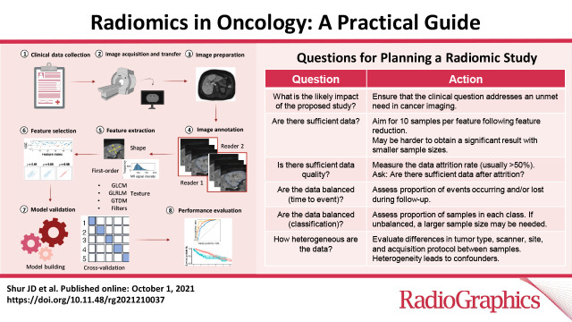
          Radiomics refers to the extraction of mineable data from medical imaging and has been applied within oncology to improve diagnosis, prognostication, and clinical decision support, with the goal of delivering precision medicine. The authors provide a practical approach for successfully implementing a radiomic workflow from planning and conceptualization through manuscript writing. Applications in oncology typically are either classification tasks that involve computing the probability of a sample belonging to a category, such as benign versus malignant, or prediction of clinical events with a time-to-event analysis, such as overall survival. The radiomic workflow is multidisciplinary, involving radiologists and data and imaging scientists, and follows a stepwise process involving tumor segmentation, image preprocessing, feature extraction, model development, and validation. Images are curated and processed before segmentation, which can be performed on tumors, tumor subregions, or peritumoral zones. Extracted features typically describe the distribution of signal intensities and spatial relationship of pixels within a region of interest. To improve model performance and reduce overfitting, redundant and nonreproducible features are removed. Validation is essential to estimate model performance in new data and can be performed iteratively on samples of the dataset (cross-validation) or on a separate hold-out dataset by using internal or external data. A variety of noncommercial and commercial radiomic software applications can be used. Guidelines and artificial intelligence checklists are useful when planning and writing up radiomic studies. Although interest in the field continues to grow, radiologists should be familiar with potential pitfalls to ensure that meaningful conclusions can be drawn.

          Online supplemental material is available for this article.

          Published under a CC BY 4.0 license.

          Abstract

          Created with BioRender.com

          Related collections

          Most cited references59

          • Record: found
          • Abstract: not found
          • Article: not found

          Controlling the False Discovery Rate: A Practical and Powerful Approach to Multiple Testing

            Bookmark
            • Record: found
            • Abstract: not found
            • Article: not found

            Regression Shrinkage and Selection Via the Lasso

              Bookmark
              • Record: found
              • Abstract: found
              • Article: not found

              Radiomics: Images Are More than Pictures, They Are Data

              This report describes the process of radiomics, its challenges, and its potential power to facilitate better clinical decision making, particularly in the care of patients with cancer.
                Bookmark

                Author and article information

                Contributors
                Journal
                Radiographics
                Radiographics
                Radiographics
                Radiographics
                Radiological Society of North America
                0271-5333
                1527-1323
                01 October 2021
                October 2021
                1 October 2022
                : 41
                : 6
                : 1717-1732
                Affiliations
                [1]From the Department of Radiology, Royal Marsden Hospital NHS Foundation Trust, Sutton, England (J.D.S., D.a.D., K.D., N. P., C.M., D.M.K.); Institute of Cancer Research, 15 Cotswold Road, Sutton SM2 5NG, England (S.J.D., S.K., J.P.B.O., N. P., C.M., D.M.K., M.R.O.); and Computational Clinical Imaging Group, Champalimaud Foundation, Centre for the Unknown, Lisbon, Portugal (N.P.).
                Author notes
                Address correspondence to M.R.O. (e-mail: matthew.orton@ 123456icr.ac.uk ).
                Author information
                http://orcid.org/0000-0002-4569-4785
                http://orcid.org/0000-0001-8569-9188
                http://orcid.org/0000-0002-6362-0616
                http://orcid.org/0000-0002-2498-4257
                http://orcid.org/0000-0003-3298-2072
                http://orcid.org/0000-0002-0557-9379
                Article
                210037
                10.1148/rg.2021210037
                8501897
                34597235
                3a7fd0ba-cbc7-4be1-b355-dae398c1ec6f
                2021 by the Radiological Society of North America, Inc.

                Published under a https://creativecommons.org/licenses/by/4.0/CC BY 4.0 license.

                History
                : 15 February 2021
                : 15 June 2021
                : 27 June 2021
                : 29 June 2021
                Funding
                Funded by: NIHR Biomedical Research Centre, Royal Marsden NHS Foundation Trust/Institute of Cancer Research http://dx.doi.org/10.13039/100014461
                Categories
                Informatics
                AI, Artificial Intelligence
                OI, Oncologic Imaging
                Custom metadata
                yes
                2024-10-01

                Comments

                Comment on this article