24
views
0
recommends
+1 Recommend
0 collections
    0
    shares
      • Record: found
      • Abstract: found
      • Article: found
      Is Open Access

      Mutation in ATG5 reduces autophagy and leads to ataxia with developmental delay

      research-article

      Read this article at

      Bookmark
          There is no author summary for this article yet. Authors can add summaries to their articles on ScienceOpen to make them more accessible to a non-specialist audience.

          Abstract

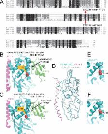
          Autophagy is required for the homeostasis of cellular material and is proposed to be involved in many aspects of health. Defects in the autophagy pathway have been observed in neurodegenerative disorders; however, no genetically-inherited pathogenic mutations in any of the core autophagy-related ( ATG) genes have been reported in human patients to date. We identified a homozygous missense mutation, changing a conserved amino acid, in ATG5 in two siblings with congenital ataxia, mental retardation, and developmental delay. The subjects' cells display a decrease in autophagy flux and defects in conjugation of ATG12 to ATG5. The homologous mutation in yeast demonstrates a 30-50% reduction of induced autophagy. Flies in which Atg5 is substituted with the mutant human ATG5 exhibit severe movement disorder, in contrast to flies expressing the wild-type human protein. Our results demonstrate the critical role of autophagy in preventing neurological diseases and maintaining neuronal health.

          DOI: http://dx.doi.org/10.7554/eLife.12245.001

          eLife digest

          Ataxia is a rare disease that affects balance and co-ordination, leading to difficulties in walking and other movements. The disease mostly affects adults, but some children are born with it and they often have additional cognitive and developmental problems. Mutations in at least 60 genes are known to be able to cause ataxia, but it is thought that there are still more to be found.

          Kim, Sandford et al. studied two siblings with the childhood form of ataxia and found that they both had a mutation in a gene called ATG5. The protein produced by the mutant ATG5 gene was less able to interact with another protein called ATG12. Furthermore, the cells of both children had defects in a process called autophagy – which destroys old and faulty proteins to prevent them accumulating and causing damage to the cell.

          Next, Kim, Sandford et al. examined the effect of this mutation in baker’s yeast cells. Cells with a mutation in the yeast equivalent of human ATG5 had lower levels of autophagy than normal cells. Further experiments used fruit flies that lacked fly Atg5, which were unable to fly or walk properly. Inserting the normal form of human ATG5 into the flies restored normal movement, but the mutant form of the gene had less of an effect.

          These findings suggest that a mutation in ATG5 can be responsible for the symptoms of childhood ataxia. Kim, Sandford et al. think that other people with severe ataxia may have mutations in genes involved in autophagy. Therefore, the next step is to study autophagy in cells from many other ataxia patients.

          DOI: http://dx.doi.org/10.7554/eLife.12245.002

          Related collections

          Most cited references37

          • Record: found
          • Abstract: found
          • Article: not found

          An optimized transgenesis system for Drosophila using germ-line-specific phiC31 integrases.

          Germ-line transformation via transposable elements is a powerful tool to study gene function in Drosophila melanogaster. However, some inherent characteristics of transposon-mediated transgenesis limit its use for transgene analysis. Here, we circumvent these limitations by optimizing a phiC31-based integration system. We generated a collection of lines with precisely mapped attP sites that allow the insertion of transgenes into many different predetermined intergenic locations throughout the fly genome. By using regulatory elements of the nanos and vasa genes, we established endogenous sources of the phiC31 integrase, eliminating the difficulties of coinjecting integrase mRNA and raising the transformation efficiency. Moreover, to discriminate between specific and rare nonspecific integration events, a white gene-based reconstitution system was generated that enables visual selection for precise attP targeting. Finally, we demonstrate that our chromosomal attP sites can be modified in situ, extending their scope while retaining their properties as landing sites. The efficiency, ease-of-use, and versatility obtained here with the phiC31-based integration system represents an important advance in transgenesis and opens up the possibility of systematic, high-throughput screening of large cDNA sets and regulatory elements.
            Bookmark
            • Record: found
            • Abstract: found
            • Article: not found

            Trehalose, a novel mTOR-independent autophagy enhancer, accelerates the clearance of mutant huntingtin and alpha-synuclein.

            Trehalose, a disaccharide present in many non-mammalian species, protects cells against various environmental stresses. Whereas some of the protective effects may be explained by its chemical chaperone properties, its actions are largely unknown. Here we report a novel function of trehalose as an mTOR-independent autophagy activator. Trehalose-induced autophagy enhanced the clearance of autophagy substrates like mutant huntingtin and the A30P and A53T mutants of alpha-synuclein, associated with Huntington disease (HD) and Parkinson disease (PD), respectively. Furthermore, trehalose and mTOR inhibition by rapamycin together exerted an additive effect on the clearance of these aggregate-prone proteins because of increased autophagic activity. By inducing autophagy, we showed that trehalose also protects cells against subsequent pro-apoptotic insults via the mitochondrial pathway. The dual protective properties of trehalose (as an inducer of autophagy and chemical chaperone) and the combinatorial strategy with rapamycin may be relevant to the treatment of HD and related diseases, where the mutant proteins are autophagy substrates.
              Bookmark
              • Record: found
              • Abstract: found
              • Article: not found

              The Atg16L complex specifies the site of LC3 lipidation for membrane biogenesis in autophagy.

              Two ubiquitin-like molecules, Atg12 and LC3/Atg8, are involved in autophagosome biogenesis. Atg12 is conjugated to Atg5 and forms an approximately 800-kDa protein complex with Atg16L (referred to as Atg16L complex). LC3/Atg8 is conjugated to phosphatidylethanolamine and is associated with autophagosome formation, perhaps by enabling membrane elongation. Although the Atg16L complex is required for efficient LC3 lipidation, its role is unknown. Here, we show that overexpression of Atg12 or Atg16L inhibits autophagosome formation. Mechanistically, the site of LC3 lipidation is determined by the membrane localization of the Atg16L complex as well as the interaction of Atg12 with Atg3, the E2 enzyme for the LC3 lipidation process. Forced localization of Atg16L to the plasma membrane enabled ectopic LC3 lipidation at that site. We propose that the Atg16L complex is a new type of E3-like enzyme that functions as a scaffold for LC3 lipidation by dynamically localizing to the putative source membranes for autophagosome formation.
                Bookmark

                Author and article information

                Contributors
                Role: Reviewing editor
                Journal
                eLife
                Elife
                eLife
                eLife
                eLife
                eLife Sciences Publications, Ltd
                2050-084X
                26 January 2016
                2016
                : 5
                : e12245
                Affiliations
                [1 ]deptDepartment of Molecular and Integrative Physiology , University of Michigan , Ann Arbor, United States
                [2 ]deptMolecular and Behavioral Neuroscience Institute , University of Michigan , Ann Arbor, United States
                [3 ]deptDepartment of Molecular, Cellular, and Developmental Biology , University of Michigan , Ann Arbor, United States
                [4 ]deptLife Sciences Institute , University of Michigan , Ann Arbor, United States
                [5 ]deptDepartment of Structural Biology , St Jude Children's Research Hospital , Memphis, United States
                [6 ]Howard Hughes Medical Institute, St. Jude Children's Research Hospital , Memphis, United States
                [7 ]deptDepartment of Human Genetics , University of Michigan , Ann Arbor, United States
                [8 ]deptDepartment of Molecular Biology and Genetics , Boğaziçi University , Istanbul, Turkey
                [9 ]deptInstitute of Genetics, Biological Research Centre , Hungarian Academy of Sciences , Szeged, Hungary
                [10 ]deptDepartment of Anatomy, Cell and Developmental Biology , Eötvös Loránd University , Budapest, Hungary
                [11 ]deptDepartment of Computational Medicine and Bioinformatics , University of Michigan , Ann Arbor, United States
                [12 ]deptDepartment of Neurology, Faculty of Medicine , Istanbul University , Istanbul, Turkey
                [13 ]deptDepartment of Psychiatry , University of Michigan , Ann Arbor, United States
                [14]The University of Tokyo , Japan
                [15]The University of Tokyo , Japan
                Author notes
                [†]

                These authors contributed equally to this work.

                Author information
                http://orcid.org/0000-0001-6727-0812
                http://orcid.org/0000-0001-8548-8874
                http://orcid.org/0000-0002-2200-6011
                http://orcid.org/0000-0002-7828-8118
                http://orcid.org/0000-0002-1914-2434
                Article
                12245
                10.7554/eLife.12245
                4786408
                26812546
                39fd66a8-195e-4d74-9657-44ae3d05d765
                © 2016, Kim et al

                This article is distributed under the terms of the Creative Commons Attribution License, which permits unrestricted use and redistribution provided that the original author and source are credited.

                History
                : 11 October 2015
                : 13 January 2016
                Funding
                Funded by: FundRef http://dx.doi.org/10.13039/100000002, National Institutes of Health;
                Award ID: OD018265
                Award Recipient :
                Funded by: FundRef http://dx.doi.org/10.13039/100000002, National Institutes of Health;
                Award ID: NS078560
                Award Recipient :
                Funded by: FundRef http://dx.doi.org/10.13039/100000968, American Heart Association;
                Award ID: 14POST19890021
                Award Recipient :
                Funded by: FundRef http://dx.doi.org/10.13039/100000011, Howard Hughes Medical Institute;
                Award Recipient :
                Funded by: FundRef http://dx.doi.org/10.13039/100000002, National Institutes of Health;
                Award ID: GM077053
                Award Recipient :
                Funded by: American Lebanese Syrian Associated Charities /St. Jude;
                Award Recipient :
                Funded by: Bogazici University Research Fund;
                Award ID: 6655
                Award Recipient :
                Funded by: FundRef http://dx.doi.org/10.13039/501100003825, Magyar Tudományos Akadémia;
                Award ID: Lendulet LP2014-2
                Award Recipient :
                Funded by: FundRef http://dx.doi.org/10.13039/100004440, Wellcome Trust;
                Award ID: 087518/Z/08/Z
                Award Recipient :
                Funded by: FundRef http://dx.doi.org/10.13039/100000002, National Institutes of Health;
                Award ID: GM053396
                Award Recipient :
                The funders had no role in study design, data collection and interpretation, or the decision to submit the work for publication.
                Categories
                Research Article
                Human Biology and Medicine
                Neuroscience
                Custom metadata
                2.5
                A genetic mutation that impairs autophagic flux leads to neurodegeneration and can cause ataxia and developmental delay in children.

                Life sciences
                ataxia,next generation sequencing,autophagy,<i>d. melanogaster</i>,human,<i>s. cerevisiae</i>

                Comments

                Comment on this article