0
views
0
recommends
+1 Recommend
0 collections
    0
    shares
      • Record: found
      • Abstract: found
      • Article: found
      Is Open Access

      A subgroup of light-driven sodium pumps with an additional Schiff base counterion

      research-article

      Read this article at

      Bookmark
          There is no author summary for this article yet. Authors can add summaries to their articles on ScienceOpen to make them more accessible to a non-specialist audience.

          Abstract

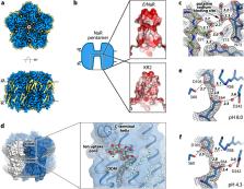
          Light-driven sodium pumps (NaRs) are unique ion-transporting microbial rhodopsins. The major group of NaRs is characterized by an NDQ motif and has two aspartic acid residues in the central region essential for sodium transport. Here we identify a subgroup of the NDQ rhodopsins bearing an additional glutamic acid residue in the close vicinity to the retinal Schiff base. We thoroughly characterize a member of this subgroup, namely the protein ErNaR from Erythrobacter sp. HL-111 and show that the additional glutamic acid results in almost complete loss of pH sensitivity for sodium-pumping activity, which is in contrast to previously studied NaRs. ErNaR is capable of transporting sodium efficiently even at acidic pH levels. X-ray crystallography and single particle cryo-electron microscopy reveal that the additional glutamic acid residue mediates the connection between the other two Schiff base counterions and strongly interacts with the aspartic acid of the characteristic NDQ motif. Hence, it reduces its pKa. Our findings shed light on a subgroup of NaRs and might serve as a basis for their rational optimization for optogenetics.

          Abstract

          Light-driven sodium-pumping rhodopsins are unique ion transporters. Here, authors present a characterization of such rhodopsins with a modified active center allowing for efficient sodium transport under various environmental conditions.

          Related collections

          Most cited references68

          • Record: found
          • Abstract: found
          • Article: found
          Is Open Access

          Highly accurate protein structure prediction with AlphaFold

          Proteins are essential to life, and understanding their structure can facilitate a mechanistic understanding of their function. Through an enormous experimental effort 1 – 4 , the structures of around 100,000 unique proteins have been determined 5 , but this represents a small fraction of the billions of known protein sequences 6 , 7 . Structural coverage is bottlenecked by the months to years of painstaking effort required to determine a single protein structure. Accurate computational approaches are needed to address this gap and to enable large-scale structural bioinformatics. Predicting the three-dimensional structure that a protein will adopt based solely on its amino acid sequence—the structure prediction component of the ‘protein folding problem’ 8 —has been an important open research problem for more than 50 years 9 . Despite recent progress 10 – 14 , existing methods fall far short of atomic accuracy, especially when no homologous structure is available. Here we provide the first computational method that can regularly predict protein structures with atomic accuracy even in cases in which no similar structure is known. We validated an entirely redesigned version of our neural network-based model, AlphaFold, in the challenging 14th Critical Assessment of protein Structure Prediction (CASP14) 15 , demonstrating accuracy competitive with experimental structures in a majority of cases and greatly outperforming other methods. Underpinning the latest version of AlphaFold is a novel machine learning approach that incorporates physical and biological knowledge about protein structure, leveraging multi-sequence alignments, into the design of the deep learning algorithm. AlphaFold predicts protein structures with an accuracy competitive with experimental structures in the majority of cases using a novel deep learning architecture.
            Bookmark
            • Record: found
            • Abstract: found
            • Article: not found

            UCSF Chimera--a visualization system for exploratory research and analysis.

            The design, implementation, and capabilities of an extensible visualization system, UCSF Chimera, are discussed. Chimera is segmented into a core that provides basic services and visualization, and extensions that provide most higher level functionality. This architecture ensures that the extension mechanism satisfies the demands of outside developers who wish to incorporate new features. Two unusual extensions are presented: Multiscale, which adds the ability to visualize large-scale molecular assemblies such as viral coats, and Collaboratory, which allows researchers to share a Chimera session interactively despite being at separate locales. Other extensions include Multalign Viewer, for showing multiple sequence alignments and associated structures; ViewDock, for screening docked ligand orientations; Movie, for replaying molecular dynamics trajectories; and Volume Viewer, for display and analysis of volumetric data. A discussion of the usage of Chimera in real-world situations is given, along with anticipated future directions. Chimera includes full user documentation, is free to academic and nonprofit users, and is available for Microsoft Windows, Linux, Apple Mac OS X, SGI IRIX, and HP Tru64 Unix from http://www.cgl.ucsf.edu/chimera/. Copyright 2004 Wiley Periodicals, Inc.
              Bookmark
              • Record: found
              • Abstract: found
              • Article: not found

              MUSCLE: multiple sequence alignment with high accuracy and high throughput.

              We describe MUSCLE, a new computer program for creating multiple alignments of protein sequences. Elements of the algorithm include fast distance estimation using kmer counting, progressive alignment using a new profile function we call the log-expectation score, and refinement using tree-dependent restricted partitioning. The speed and accuracy of MUSCLE are compared with T-Coffee, MAFFT and CLUSTALW on four test sets of reference alignments: BAliBASE, SABmark, SMART and a new benchmark, PREFAB. MUSCLE achieves the highest, or joint highest, rank in accuracy on each of these sets. Without refinement, MUSCLE achieves average accuracy statistically indistinguishable from T-Coffee and MAFFT, and is the fastest of the tested methods for large numbers of sequences, aligning 5000 sequences of average length 350 in 7 min on a current desktop computer. The MUSCLE program, source code and PREFAB test data are freely available at http://www.drive5. com/muscle.
                Bookmark

                Author and article information

                Contributors
                alexey.alekseev@med.uni-goettingen.de
                kirill.kovalev@embl-hamburg.de
                Journal
                Nat Commun
                Nat Commun
                Nature Communications
                Nature Publishing Group UK (London )
                2041-1723
                10 April 2024
                10 April 2024
                2024
                : 15
                : 3119
                Affiliations
                [1 ]Department of Ophthalmology, University Hospital Bonn, Medical Faculty, ( https://ror.org/01xnwqx93) Bonn, Germany
                [2 ]Institute of Physical and Theoretical Chemistry, Goethe University Frankfurt, ( https://ror.org/04cvxnb49) 60438 Frankfurt am Main, Germany
                [3 ]Groningen Institute for Biomolecular Sciences and Biotechnology, University of Groningen, ( https://ror.org/012p63287) 9747AG Groningen, the Netherlands
                [4 ]Fritz Haber Center for Molecular Dynamics Research, Institute of Chemistry, The Hebrew University of Jerusalem, ( https://ror.org/03qxff017) Jerusalem, 9190401 Israel
                [5 ]Department of Biochemistry, University of Cambridge, ( https://ror.org/013meh722) 80 Tennis Court Road, Cambridge, CB2 1GA UK
                [6 ]European Molecular Biology Laboratory, EMBL Hamburg c/o DESY, ( https://ror.org/03mstc592) 22607 Hamburg, Germany
                [7 ]Institute of Biological Information Processing (IBI-7: Structural Biochemistry), Forschungszentrum Jülich, ( https://ror.org/02nv7yv05) Jülich, Germany
                [8 ]JuStruct: Jülich Center for Structural Biology, Forschungszentrum Jülich, ( https://ror.org/02nv7yv05) Jülich, Germany
                [9 ]GRID grid.418192.7, ISNI 0000 0004 0641 5776, Univ. Grenoble Alpes, CEA, CNRS, , Institut de Biologie Structurale (IBS), ; 38000 Grenoble, France
                [10 ]GRID grid.52788.30, ISNI 0000 0004 0427 7672, European Molecular Biology Laboratory, European Bioinformatics Institute (EMBL-EBI), , Wellcome Genome Campus, ; Hinxton, UK
                [11 ]University Medical Center Göttingen, Institute for Auditory Neuroscience and InnerEarLab, ( https://ror.org/021ft0n22) Robert-Koch-Str. 40, 37075 Göttingen, Germany
                [12 ]Cluster of Excellence “Multiscale Bioimaging: from Molecular Machines to Networks of Excitable Cells” (MBExC), University of Göttingen, ( https://ror.org/01y9bpm73) Göttingen, Germany
                [13 ]Present Address: School of Chemistry, University of Edinburgh, ( https://ror.org/01nrxwf90) Edinburgh, EH9 3FJ UK
                Author information
                http://orcid.org/0000-0002-3560-1645
                http://orcid.org/0009-0006-6743-2344
                http://orcid.org/0000-0001-5019-2096
                http://orcid.org/0000-0003-3032-3794
                http://orcid.org/0000-0003-3181-0236
                http://orcid.org/0000-0001-8471-8041
                http://orcid.org/0000-0001-6955-7374
                http://orcid.org/0000-0002-6982-4660
                http://orcid.org/0000-0002-8496-8240
                http://orcid.org/0000-0001-8536-6869
                http://orcid.org/0000-0001-7517-8944
                http://orcid.org/0000-0003-2340-2216
                http://orcid.org/0000-0001-8990-1662
                http://orcid.org/0000-0002-2229-181X
                Article
                47469
                10.1038/s41467-024-47469-0
                11006869
                38600129
                398fdd3e-5e64-4c1e-a30a-d45ff84b3390
                © The Author(s) 2024

                Open Access This article is licensed under a Creative Commons Attribution 4.0 International License, which permits use, sharing, adaptation, distribution and reproduction in any medium or format, as long as you give appropriate credit to the original author(s) and the source, provide a link to the Creative Commons licence, and indicate if changes were made. The images or other third party material in this article are included in the article’s Creative Commons licence, unless indicated otherwise in a credit line to the material. If material is not included in the article’s Creative Commons licence and your intended use is not permitted by statutory regulation or exceeds the permitted use, you will need to obtain permission directly from the copyright holder. To view a copy of this licence, visit http://creativecommons.org/licenses/by/4.0/.

                History
                : 12 October 2023
                : 1 April 2024
                Funding
                Funded by: FundRef https://doi.org/10.13039/100010665, EC | EU Framework Programme for Research and Innovation H2020 | H2020 Priority Excellent Science | H2020 Marie Skłodowska-Curie Actions (H2020 Excellent Science - Marie Skłodowska-Curie Actions);
                Award ID: 847543
                Award Recipient :
                Categories
                Article
                Custom metadata
                © Springer Nature Limited 2024

                Uncategorized
                permeation and transport,x-ray crystallography,cryoelectron microscopy
                Uncategorized
                permeation and transport, x-ray crystallography, cryoelectron microscopy

                Comments

                Comment on this article