1
views
0
recommends
+1 Recommend
0 collections
    0
    shares
      • Record: found
      • Abstract: found
      • Article: found
      Is Open Access

      Integrated Design and Prototyping of a Robotic Head for Ocular and Craniofacial Trauma Simulators

      research-article

      Read this article at

      Bookmark
          There is no author summary for this article yet. Authors can add summaries to their articles on ScienceOpen to make them more accessible to a non-specialist audience.

          ABSTRACT

          Background

          Medical simulation is relevant for training medical personnel in the delivery of medical and trauma care, with benefits including quantitative evaluation and increased patient safety through reduced need to train on patients.

          Methods

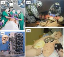
          This paper presents a prototype medical simulator focusing on ocular and craniofacial trauma (OCF), for training in management of facial and upper airway injuries. It consists of a physical, electromechanical representation of head and neck structures, including the mandible, maxillary region, neck, orbit and peri‐orbital regions to replicate different craniofacial traumas. Actuation and hydraulic systems are designed to control animatronic features and flow of simulated blood, tears, and cerebrospinal fluid.

          Results

          Experimentally validated, the OCF simulator achieves structural and functional characteristics as close as possible to those of a human body.

          Conclusions

          The OCF Simulator can be used as a stand‐alone active simulator, it can be transported and used to train surgeons in simulated real‐life scenarios.

          Clinical Trial Registration

          The authors declare that this statement is not applicable since no clinical tests have been performed.

          Related collections

          Most cited references30

          • Record: found
          • Abstract: found
          • Article: not found

          Simulation based medical education: an opportunity to learn from errors.

          Medical professionals and educators recognize that Simulation Based Medical Education (SBME) can contribute considerably to improving medical care by boosting medical professionals' performance and enhancing patient safety. A central characteristic of SBME is its unique approach to making (and learning from) mistakes, which is regarded as a powerful educational experience and as an opportunity for professional improvement. The basic assumption underlying SBME is that increased practice in learning from mistakes and in error management in a simulated environment will reduce occurrences of errors in real life and will provide professionals with the correct attitude and skills to cope competently with those mistakes that could not be prevented. The main message of the present paper is that this assumption, which serves as the driving force of SBME, should also serve as a starting point for critical thinking and questioning regarding the multiple aspects and components of SBME. These questions, in turn, should lead to empirical research that will provide feedback concerning changes that may be necessary in order to attain the goal of improving medical professionals' performance. Based on such research, SBME will be held accountable for its outcomes, i.e. whether its educational techniques indeed result in decreased occurrence of errors or not, and whether the ability to cope with the errors that do occur is significantly improved. The first of three issues that were addressed concerns individuals' experience of performing mistakes. It is suggested that in order to benefit fully from the experience of performing mistakes in a simulated context, medical educators should create a balance between the emotional load associated with the experience and the professional lessons that can be learned. Furthermore, research should focus on the long-term effects of the experience in changing professionals' attitudes and behaviour. The second question concerned the contribution of the different components of the educational experience to creating the desired changes in professionals' performance. Analysis of the teaching and learning involved in each stage of the educational event should serve as the basis for research that aims at identifying the unique contribution and efficiency of each element, and defining the essential core activities of a simulated experience. Finally, the need to define a newly emerging profession-SBME educator-was addressed. The professional qualifications are, clearly, multidisciplinary and should be based on the growing experience of medical educators in training students and professionals. Defining the profession is essential in order to create academic environments in which professionals will be trained to develop and implement new programmes, accompanied by research and assessment.
            Bookmark
            • Record: found
            • Abstract: found
            • Article: not found

            Carotid artery diameter in men and women and the relation to body and neck size.

            To explore relationships among gender, body size, neck size, and the diameters of the common carotid artery (CCA) and internal carotid artery (ICA). Using multivariate regression, the best predictors of sonographic diameters of CCA and ICA were determined based on age, height, weight, body mass index, body surface area, neck circumference, neck length, and blood pressure. Measurements were obtained in 500 consecutive patients (age 52+/-15 years; 61% women). Mean diameters of ICA (4.66+/-0.78 mm) and CCA (6.10+/-0.80 mm) in women were significantly smaller than in men: 5.11+/-0.87 mm and 6.52+/-0.98 mm, respectively. Sex significantly influenced the diameters after controlling for body size, neck size, age, and blood pressure. Carotid arteries are smaller in women even after adjusting for body and neck size, age, and blood pressure.
              Bookmark
              • Record: found
              • Abstract: found
              • Article: not found

              Anatomy of the temporomandibular joint.

              The temporomandibular joint (TMJ), also known as the mandibular joint, is an ellipsoid variety of the right and left synovial joints forming a bicondylar articulation. The common features of the synovial joints exhibited by this joint include a fibrous capsule, a disk, synovial membrane, fluid, and tough adjacent ligaments. Not only is the mandible a single bone but the cranium is also mechanically a single stable component; therefore, the correct terminology for the joint is the craniomandibular articulation. The term temporomandibular joint is misleading and seems to only refer to one side when referring to joint function. Magnetic resonance imaging has been shown to accurately delineate the structures of the TMJ and is the best technique to correlate and compare the TMJ components such as bone, disk, fluid, capsule, and ligaments with autopsy specimens.
                Bookmark

                Author and article information

                Contributors
                roberto.pitzalis@iit.it
                Journal
                Int J Med Robot
                Int J Med Robot
                10.1002/(ISSN)1478-596X
                RCS
                The International Journal of Medical Robotics + Computer Assisted Surgery
                John Wiley and Sons Inc. (Hoboken )
                1478-5951
                1478-596X
                02 January 2025
                February 2025
                : 21
                : 1 ( doiID: 10.1002/rcs.v21.1 )
                : e70039
                Affiliations
                [ 1 ] Department of Imaging Massachusetts General Hospital and Harvard Medical School Cambridge Massachusetts USA
                [ 2 ] Department of Mechanical Energy Management and Transportation Engineering University of Genova Genova Italy
                [ 3 ] ADVR Department Istituto Italiano di Tecnologia Genova Italy
                Author notes
                [*] [* ] Correspondence: Roberto F. Pitzalis

                ( roberto.pitzalis@ 123456iit.it )

                Author information
                https://orcid.org/0000-0003-1464-6219
                Article
                RCS70039
                10.1002/rcs.70039
                11694338
                39744974
                39150587-eb06-44e8-bd8e-596d234a0508
                © 2025 The Author(s). The International Journal of Medical Robotics and Computer Assisted Surgery published by John Wiley & Sons Ltd.

                This is an open access article under the terms of the http://creativecommons.org/licenses/by-nc/4.0/ License, which permits use, distribution and reproduction in any medium, provided the original work is properly cited and is not used for commercial purposes.

                History
                : 06 December 2024
                : 12 July 2024
                : 17 December 2024
                Page count
                Figures: 22, Tables: 4, Pages: 14, Words: 8390
                Funding
                Funded by: US Department of the Army
                Award ID: W81XWH‐11‐C‐0095
                Categories
                Original Article
                Original Article
                Custom metadata
                2.0
                February 2025
                Converter:WILEY_ML3GV2_TO_JATSPMC version:6.5.1 mode:remove_FC converted:02.01.2025

                Surgery
                craniofacial trauma,medical simulators,physician training,robotic human head
                Surgery
                craniofacial trauma, medical simulators, physician training, robotic human head

                Comments

                Comment on this article

                scite_
                0
                0
                0
                0
                Smart Citations
                0
                0
                0
                0
                Citing PublicationsSupportingMentioningContrasting
                View Citations

                See how this article has been cited at scite.ai

                scite shows how a scientific paper has been cited by providing the context of the citation, a classification describing whether it supports, mentions, or contrasts the cited claim, and a label indicating in which section the citation was made.

                Similar content59

                Most referenced authors202