37
views
0
recommends
+1 Recommend
0 collections
    0
    shares
      • Record: found
      • Abstract: found
      • Article: found
      Is Open Access

      Nrf2 drives oxidative stress-induced autophagy in nucleus pulposus cells via a Keap1/Nrf2/p62 feedback loop to protect intervertebral disc from degeneration

      research-article

      Read this article at

      Bookmark
          There is no author summary for this article yet. Authors can add summaries to their articles on ScienceOpen to make them more accessible to a non-specialist audience.

          Abstract

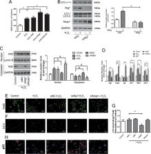
          Intervertebral disc (IVD) degeneration is known to aggravate with age and oxidative stress is implicated in the pathogenesis of many age-related diseases. Nuclear factor (erythroid-derived-2)-like 2 (Nrf2) can confer adaptive protection against oxidative and proteotoxic stress in cells. In this study, we assessed whether Nrf2 can protect against oxidative stress in nucleus pulposus (NP) cells. In addition, we investigated Nrf2 expression in NP tissue samples from patients with different degrees of IVD degeneration and a mouse model of aging and IVD degeneration and the influence of H 2O 2-induced oxidative stress on autophagic pathways in NP cells. Autophagy was assessed by measuring levels of autophagy-related protein (ATG) family members and the autophagic markers, p62 and LC3. We found that expression of Nrf2 progressively decreased in human NP tissue samples of patients with increasing degrees of IVD degeneration. Nrf2 deficiency leads to the degeneration of IVDs during aging. Nrf2 knockout also aggravates IVD degeneration and reduces autophagic gene expression in an induced mouse model of IVD degeneration. The detrimental effects of H 2O 2-induced oxidative stress were increased in autophagy-deficient cells via reduced expression of Atg7 and the Keap1–Nrf2–p62 autophagy pathway. Taken together, these results suggest that excessive oxidative stress causes the upregulation of autophagy, and autophagy acts as an antioxidant feedback response activated by a Keap1-Nrf2-p62 feedback loop in IVD degeneration.

          Related collections

          Most cited references24

          • Record: found
          • Abstract: found
          • Article: not found

          Molecular mechanisms of the Keap1–Nrf2 pathway in stress response and cancer evolution.

          The Keap1–Nrf2 regulatory pathway plays a central role in the protection of cells against oxidative and xenobiotic damage. Under unstressed conditions, Nrf2 is constantly ubiquitinated by the Cul3–Keap1 ubiquitin E3 ligase complex and rapidly degraded in proteasomes. Upon exposure to electrophilic and oxidative stresses, reactive cysteine residues of Keap1 become modified, leading to a decline in the E3 ligase activity, stabilization of Nrf2 and robust induction of a battery of cytoprotective genes. Biochemical and structural analyses have revealed that the intact Keap1 homodimer forms a cherry-bob structure in which one molecule of Nrf2 associates with two molecules of Keap1 by using two binding sites within the Neh2 domain of Nrf2. This two-site binding appears critical for Nrf2 ubiquitination. In many human cancers, missense mutations in KEAP1 and NRF2 genes have been identified. These mutations disrupt the Keap1–Nrf2 complex activity involved in ubiquitination and degradation of Nrf2 and result in constitutive activation of Nrf2. Elevated expression of Nrf2 target genes confers advantages in terms of stress resistance and cell proliferation in normal and cancer cells. Discovery and development of selective Nrf2 inhibitors should make a critical contribution to improved cancer therapy.
            Bookmark
            • Record: found
            • Abstract: found
            • Article: not found

            The Atg12-Atg5 conjugate has a novel E3-like activity for protein lipidation in autophagy.

            Autophagy is a bulk degradation process in eukaryotic cells; autophagosomes enclose cytoplasmic components for degradation in the lysosome/vacuole. Autophagosome formation requires two ubiquitin-like conjugation systems, the Atg12 and Atg8 systems, which are tightly associated with expansion of autophagosomal membrane. Previous studies have suggested that there is a hierarchy between these systems; the Atg12 system is located upstream of the Atg8 system in the context of Atg protein organization. However, the concrete molecular relationship is unclear. Here, we show using an in vitro Atg8 conjugation system that the Atg12-Atg5 conjugate, but not unconjugated Atg12 or Atg5, strongly enhances the formation of the other conjugate, Atg8-PE. The Atg12-Atg5 conjugate promotes the transfer of Atg8 from Atg3 to the substrate, phosphatidylethanolamine (PE), by stimulating the activity of Atg3. We also show that the Atg12-Atg5 conjugate interacts with both Atg3 and PE-containing liposomes. These results indicate that the Atg12-Atg5 conjugate is a ubiquitin-protein ligase (E3)-like enzyme for Atg8-PE conjugation reaction, distinctively promoting protein-lipid conjugation.
              Bookmark
              • Record: found
              • Abstract: found
              • Article: not found

              Degeneration of the intervertebral disc

              The intervertebral disc is a cartilaginous structure that resembles articular cartilage in its biochemistry, but morphologically it is clearly different. It shows degenerative and ageing changes earlier than does any other connective tissue in the body. It is believed to be important clinically because there is an association of disc degeneration with back pain. Current treatments are predominantly conservative or, less commonly, surgical; in many cases there is no clear diagnosis and therapy is considered inadequate. New developments, such as genetic and biological approaches, may allow better diagnosis and treatments in the future.
                Bookmark

                Author and article information

                Contributors
                +(86) 021-81885621 , spine.chen@163.com
                Journal
                Cell Death Dis
                Cell Death Dis
                Cell Death & Disease
                Nature Publishing Group UK (London )
                2041-4889
                1 July 2019
                1 July 2019
                July 2019
                : 10
                : 7
                : 510
                Affiliations
                ISNI 0000 0004 0369 1660, GRID grid.73113.37, Department of Spinal Surgery, Changzheng Hospital, , Second Military Medical University, ; Shanghai, China
                Article
                1701
                10.1038/s41419-019-1701-3
                6602960
                31263165
                38bf8b59-1792-485e-8d3a-235f4c32ea58
                © The Author(s) 2019

                Open Access This article is licensed under a Creative Commons Attribution 4.0 International License, which permits use, sharing, adaptation, distribution and reproduction in any medium or format, as long as you give appropriate credit to the original author(s) and the source, provide a link to the Creative Commons license, and indicate if changes were made. The images or other third party material in this article are included in the article’s Creative Commons license, unless indicated otherwise in a credit line to the material. If material is not included in the article’s Creative Commons license and your intended use is not permitted by statutory regulation or exceeds the permitted use, you will need to obtain permission directly from the copyright holder. To view a copy of this license, visit http://creativecommons.org/licenses/by/4.0/.

                History
                : 26 October 2018
                : 28 May 2019
                : 29 May 2019
                Categories
                Article
                Custom metadata
                © The Author(s) 2019

                Cell biology
                autophagy,cell signalling
                Cell biology
                autophagy, cell signalling

                Comments

                Comment on this article