3
views
0
recommends
+1 Recommend
0 collections
    0
    shares
      • Record: found
      • Abstract: found
      • Article: found
      Is Open Access

      The Association of Retinal Microvasculature With Gray Matter Changes and Structural Covariance Network: A Voxel-Based Morphometry Study

      research-article

      Read this article at

      Bookmark
          There is no author summary for this article yet. Authors can add summaries to their articles on ScienceOpen to make them more accessible to a non-specialist audience.

          Abstract

          Purpose

          Increasing evidence suggests that retinal microvasculature may reflect global cerebral atrophy. However, little is known about the relation of retinal microvasculature with specific brain regions and brain networks. Therefore, we aimed to unravel the association of retinal microvasculature with gray matter changes and structural covariance network using a voxel-based morphometry (VBM) analysis.

          Methods

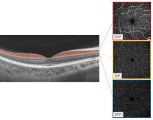
          One hundred and forty-four volunteers without previously known neurological diseases were recruited from West China Hospital, Sichuan University between April 1, 2021, and December 31, 2021. Retinal microvasculature of superficial vascular plexus (SVP), intermediate capillary plexus (ICP), and deep capillary plexus (DCP) were measured by optical coherence tomography angiography using an automatic segmentation. The VBM and structural covariance network analyses were applied to process brain magnetic resonance imaging (MRI) images. The associations of retinal microvasculature with voxel-wise gray matter volumes and structural covariance network were assessed by linear regression models.

          Results

          In the study, 137 participants (mean age = 59.72 years, 37.2% men) were included for the final analysis. Reduced perfusion in SVP was significantly associated with reduced voxel-wise gray matter volumes of the brain regions including the insula, putamen, occipital, frontal, and temporal lobes, all of which were located in the anterior part of the brain supplied by internal carotid artery, except the occipital lobe. In addition, these regions were also involved in visual processing and cognitive impairment (such as left inferior occipital gyrus, left lingual gyrus, and right parahippocampal gyrus). In regard to the structural covariance, the perfusions in SVP were positively related to the structural covariance of the left lingual gyrus seed with the left middle occipital gyrus, the right middle occipital gyrus, and the left middle frontal gyrus.

          Conclusions

          Poor perfusion in SVP was correlated with reduced voxel-wise gray matter volumes and structural covariance networks in regions related to visual processing and cognitive impairment. It suggests that retinal microvasculature may offer a window to identify aging related cerebral alterations.

          Related collections

          Most cited references56

          • Record: found
          • Abstract: found
          • Article: found

          Neuroimaging standards for research into small vessel disease and its contribution to ageing and neurodegeneration

          Summary Cerebral small vessel disease (SVD) is a common accompaniment of ageing. Features seen on neuroimaging include recent small subcortical infarcts, lacunes, white matter hyperintensities, perivascular spaces, microbleeds, and brain atrophy. SVD can present as a stroke or cognitive decline, or can have few or no symptoms. SVD frequently coexists with neurodegenerative disease, and can exacerbate cognitive deficits, physical disabilities, and other symptoms of neurodegeneration. Terminology and definitions for imaging the features of SVD vary widely, which is also true for protocols for image acquisition and image analysis. This lack of consistency hampers progress in identifying the contribution of SVD to the pathophysiology and clinical features of common neurodegenerative diseases. We are an international working group from the Centres of Excellence in Neurodegeneration. We completed a structured process to develop definitions and imaging standards for markers and consequences of SVD. We aimed to achieve the following: first, to provide a common advisory about terms and definitions for features visible on MRI; second, to suggest minimum standards for image acquisition and analysis; third, to agree on standards for scientific reporting of changes related to SVD on neuroimaging; and fourth, to review emerging imaging methods for detection and quantification of preclinical manifestations of SVD. Our findings and recommendations apply to research studies, and can be used in the clinical setting to standardise image interpretation, acquisition, and reporting. This Position Paper summarises the main outcomes of this international effort to provide the STandards for ReportIng Vascular changes on nEuroimaging (STRIVE).
            Bookmark
            • Record: found
            • Abstract: found
            • Article: not found

            MR signal abnormalities at 1.5 T in Alzheimer's dementia and normal aging.

            The type, frequency, and extent of MR signal abnormalities in Alzheimer's disease and normal aging are a subject of controversy. With a 1.5-MR unit we studied 12 Alzheimer patients, four subjects suffering from multiinfarct dementia and nine age-matched controls. Punctate or early confluent high-signal abnormalities in the deep white matter, noted in 60% of both Alzheimer patients and controls, were unrelated to the presence of hypertension or other vascular risk factors. A significant number of Alzheimer patients exhibited a more extensive smooth "halo" of periventricular hyperintensity when compared with controls (p = .024). Widespread deep white-matter hyperintensity (two patients) and extensive, irregular periventricular hyperintensity (three patients) were seen in multiinfarct dementia. Areas of high signal intensity affecting hippocampal and sylvian cortex were also present in five Alzheimer and two multiinfarct dementia patients, but absent in controls. Discrete, small foci of deep white-matter hyperintensity are not characteristic of Alzheimer's disease nor do they appear to imply a vascular cause for the dementing illness. The frequently observed "halo" of periventricular hyperintensity in Alzheimer's disease may be of diagnostic importance. High-signal abnormalities in specific cortical regions are likely to reflect disease processes localized to those structures.
              Bookmark
              • Record: found
              • Abstract: found
              • Article: not found

              Hemispheric asymmetry reduction in older adults: the HAROLD model.

              A model of the effects of aging on brain activity during cognitive performance is introduced. The model is called HAROLD (hemispheric asymmetry reduction in older adults), and it states that, under similar circumstances, prefrontal activity during cognitive performances tends to be less lateralized in older adults than in younger adults. The model is supported by functional neuroimaging and other evidence in the domains of episodic memory, semantic memory, working memory, perception, and inhibitory control. Age-related hemispheric asymmetry reductions may have a compensatory function or they may reflect a dedifferentiation process. They may have a cognitive or neural origin, and they may reflect regional or network mechanisms. The HAROLD model is a cognitive neuroscience model that integrates ideas and findings from psychology and neuroscience of aging.
                Bookmark

                Author and article information

                Journal
                Invest Ophthalmol Vis Sci
                Invest Ophthalmol Vis Sci
                IOVS
                Investigative Ophthalmology & Visual Science
                The Association for Research in Vision and Ophthalmology
                0146-0404
                1552-5783
                28 December 2023
                December 2023
                : 64
                : 15
                : 40
                Affiliations
                [1 ]Department of Neurology, West China Hospital, Sichuan University China, Chengdu, Sichuan Province, China
                [2 ]School of Information Science and Engineering, Yanshan University, Qinhuangdao, China
                [3 ]Department of Radiology, West China Hospital, Sichuan University China, Chengdu, Sichuan Province, China
                Author notes
                [# ]Correspondence: Ming Liu, Centre of Cerebrovascular Diseases, Department of Neurology, West China Hospital, Sichuan University, No. 37, Guo Xue Xiang, Chengdu 610041, China; wyplmh@ 123456hotmail.com .
                Xiaonan Guo, School of Information Science and Engineering, Yanshan University, Qinhuangdao 066104, China; guoxiaonan@ 123456ysu.edu.cn .
                [*]

                JL and WT contributed equally to this work.

                Article
                IOVS-23-37525
                10.1167/iovs.64.15.40
                10756243
                38153752
                37e1dfe0-5f14-4a35-bdac-ef52fb4011db
                Copyright 2023 The Authors

                This work is licensed under a Creative Commons Attribution-NonCommercial-NoDerivatives 4.0 International License.

                History
                : 28 November 2023
                : 26 April 2023
                Page count
                Pages: 9
                Categories
                Visual Neuroscience
                Visual Neuroscience

                retinal microvasculature,superficial vascular plexus (svp),intermediate capillary plexus (icp),deep capillary plexus (dcp),structural covariance,voxel-based morphometry analysis

                Comments

                Comment on this article