6
views
0
recommends
+1 Recommend
0 collections
    0
    shares
      • Record: found
      • Abstract: found
      • Article: found
      Is Open Access

      Potential role of post-treatment follow-up FDG-PET CT to detect mandibular osteoradionecrosis: A case report

      research-article

      Read this article at

      Bookmark
          There is no author summary for this article yet. Authors can add summaries to their articles on ScienceOpen to make them more accessible to a non-specialist audience.

          Abstract

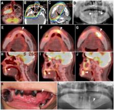
          It was hypothesized that fluorodeoxyglucose (FDG) uptake on post-treatment follow-up positron emission tomography with computed tomography (PET CT; using PET CT to monitor and rule out recurrence and metastasis of head and neck carcinoma) would be useful for detecting and understanding the disease state of osteoradionecrosis (ORN) of the jaw. The present study included 14 patients who developed mandibular ORN following radiation therapy (RT) for head and neck cancer and underwent follow-up PET CT several times following RT. Areas exhibiting FDG uptake were retrospectively assessed on post-treatment follow-up PET CT images and were classified into three types: Spot type: Only spot accumulation of FDG; localized type: Accumulation of FDG restricted to within the bone resorption area; extensive type: Accumulation of FDG extending into surrounding soft tissue. PET classification at the time of clinical diagnosis of mandibular ORN in the 14 patients demonstrated the extensive type in 43%, localized type in 36% and spot type in 21%. An increased area of FDG uptake around the ORN was revealed retrospectively on post-treatment follow-up FDG PET-CT images in 50% of patients. Alterations in PET classification included spot type to localized type in 36% and localized type to extensive type in 14%. A significantly increased number of patients with extensive-type ORN (P=0.026) required surgery. Post-treatment follow-up FDG-PET CT may be useful for early detection and better understanding of ORN.

          Related collections

          Most cited references15

          • Record: found
          • Abstract: found
          • Article: not found

          Incidence and prevention of osteoradionecrosis after dental extraction in irradiated patients: a systematic review.

          This systematic review aims to identify and review the best available evidence to answer the clinical question 'What are the incidence and the factors influencing the development of osteoradionecrosis after tooth extraction in irradiated patients?'. A systematic review of published articles on post-irradiation extraction was performed via electronic search of the Medline, Ovid, Embase and Cochrane Library databases. Additional studies were identified by manual reference list search. Evaluation and critical appraisal were done in 3 stages by two independent reviewers and any disagreement was resolved by discussion with a third party. 19 articles were selected for the final analysis. The total incidence of osteoradionecrosis after tooth extraction in irradiated patients was 7%. When extractions were performed in conjunction with prophylactic hyperbaric oxygen, the incidence was 4% while extraction in conjunction with antibiotics gave an incidence of 6%. This systematic review found that while the incidence of osteoradionecrosis after post-irradiation tooth extractions is low, the extraction of mandibular teeth within the radiation field in patients who received a radiation dose higher than 60Gy represents the highest risk of developing osteoradionecrosis. Based on weak evidence, prophylactic hyperbaric oxygen is effective in reducing the risk of developing osteoradionecrosis after post-radiation extractions. Copyright © 2010 International Association of Oral and Maxillofacial Surgeons. Published by Elsevier Ltd. All rights reserved.
            Bookmark
            • Record: found
            • Abstract: found
            • Article: not found

            Hyperbaric oxygen therapy for radionecrosis of the jaw: a randomized, placebo-controlled, double-blind trial from the ORN96 study group.

            To determine the efficacy and safety of hyperbaric oxygen therapy (HBO) for overt mandibular osteoradionecrosis. This prospective, multicenter, randomized, double-blind, placebo-controlled trial was conducted at 12 university hospitals. Ambulatory adults with overt osteoradionecrosis of the mandible were assigned to receive 30 HBO exposures preoperatively at 2.4 absolute atmosphere for 90 minutes or a placebo, and 10 additional HBO dives postoperatively or a placebo. The main outcome measure was 1-year recovery rate from osteoradionecrosis. Secondary end points included time to treatment failure, time to pain relief, 1-year mortality rate, and treatment safety. At the time of the second interim analysis, based on the triangular test, the study was stopped for potentially worse outcomes in the HBO arm. A total of 68 patients were enrolled and analyzed. At 1 year, six (19%) of 31 patients had recovered in the HBO arm and 12 (32%) of 37 in the placebo arm (relative risk = 0.60; 95% CI, 0.25 to 1.41; P = .23). Time to treatment failure (hazard ratio = 1.33; 95% CI, 0.68 to 2.60; P = .41) and time to pain relief (hazard ratio = 1.00; 95% CI, 0.52 to 1.89; P = .99) were similar between the two treatment arms. Patients with overt mandibular osteoradionecrosis did not benefit from hyperbaric oxygenation.
              Bookmark
              • Record: found
              • Abstract: found
              • Article: not found

              Paradigm shifts in the management of osteoradionecrosis of the mandible.

              Osteoradionecrosis (ORN) of the mandible is a significant complication of radiation therapy for head and neck cancer. In this condition, bone within the radiation field becomes devitalized and exposed through the overlying skin or mucosa, persisting as a non-healing wound for three months or more. In 1926, Ewing first recognized the bone changes associated with radiation therapy and described them as "radiation osteitis". In 1983, Marx proposed the first staging system for ORN that also served as a treatment protocol. This protocol advocated that patients whose disease progressed following conservative therapy (hyperbaric oxygen (HBO), local wound care, debridement) were advanced to a radical resection with a staged reconstruction utilizing a non-vascularized bone graft. Since the introduction of Marx's protocol, there have been advances in surgical techniques (i.e. microvascular surgery), as well as in imaging techniques, which have significantly impacted on the diagnosis and management of ORN. High resolution CT scans and orthopantamograms have become a key component in evaluating and staging ORN, prior to formulating a treatment plan. Patients can now be stratified based on imaging and clinical findings, and treatment can be determined based on the stage of disease, rather than determining the stage of disease based on a patient's response to a standardized treatment protocol. Reconstructions are now routinely performed immediately after resection of the diseased tissue rather than in a staged fashion. Furthermore, the transfer of well-vascularized hard and soft tissue using microvascular surgery have brought the utility of HBO treatment in advanced ORN into question. Copyright © 2010 Elsevier Ltd. All rights reserved.
                Bookmark

                Author and article information

                Journal
                Mol Clin Oncol
                Mol Clin Oncol
                MCO
                Molecular and Clinical Oncology
                D.A. Spandidos
                2049-9450
                2049-9469
                January 2018
                02 November 2017
                02 November 2017
                : 8
                : 1
                : 61-67
                Affiliations
                [1 ]Department of Oral and Maxillofacial Surgery, Kobe University Graduate School of Medicine, Kobe 650-0017, Japan
                [2 ]Department of Plastic Surgery, Kobe University Graduate School of Medicine, Kobe 650-0017, Japan
                Author notes
                Correspondence to: Dr Masaya Akashi, Department of Oral and Maxillofacial Surgery, Kobe University Graduate School of Medicine, 7-5-2 Kusunoki-cho, Chuo-ku, Kobe 650-0017, Japan, E-mail: akashim@ 123456med.kobe-u.ac.jp
                Article
                MCO-0-0-1477
                10.3892/mco.2017.1477
                5769210
                37a6ed16-93b0-44a2-bad6-d436eb740513
                Copyright: © Akashi et al.

                This is an open access article distributed under the terms of the Creative Commons Attribution-NonCommercial-NoDerivs License, which permits use and distribution in any medium, provided the original work is properly cited, the use is non-commercial and no modifications or adaptations are made.

                History
                : 29 August 2017
                : 30 October 2017
                Categories
                Articles

                mandible,osteoradionecrosis,positron emission tomography,early detection,fluorodeoxyglucose

                Comments

                Comment on this article