8
views
0
recommends
+1 Recommend
1 collections
    0
    shares
      • Record: found
      • Abstract: found
      • Article: not found

      Mitochondrial Dynamics in SARS-COV2 Spike Protein Treated Human Microglia: Implications for Neuro-COVID

      research-article

      Read this article at

      ScienceOpenPublisherPMC
      Bookmark
          There is no author summary for this article yet. Authors can add summaries to their articles on ScienceOpen to make them more accessible to a non-specialist audience.

          Abstract

          Abstract

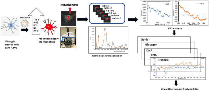
          Emerging clinical data from the current COVID-19 pandemic suggests that ~ 40% of COVID-19 patients develop neurological symptoms attributed to viral encephalitis while in COVID long haulers chronic neuro-inflammation and neuronal damage result in a syndrome described as Neuro-COVID. We hypothesize that SAR-COV2 induces mitochondrial dysfunction and activation of the mitochondrial-dependent intrinsic apoptotic pathway, resulting in microglial and neuronal apoptosis. The goal of our study was to determine the effect of SARS-COV2 on mitochondrial biogenesis and to monitor cell apoptosis in human microglia non-invasively in real time using Raman spectroscopy, providing a unique spatio-temporal information on mitochondrial function in live cells. We treated human microglia with SARS-COV2 spike protein and examined the levels of cytokines and reactive oxygen species (ROS) production, determined the effect of SARS-COV2 on mitochondrial biogenesis and examined the changes in molecular composition of phospholipids. Our results show that SARS- COV2 spike protein increases the levels of pro-inflammatory cytokines and ROS production, increases apoptosis and increases the oxygen consumption rate (OCR) in microglial cells. Increases in OCR are indicative of increased ROS production and oxidative stress suggesting that SARS-COV2 induced cell death. Raman spectroscopy yielded significant differences in phospholipids such as Phosphatidylinositol (PI), phosphatidylserine (PS), phosphatidylethanolamine (PE) and phosphatidylcholine (PC), which account for ~ 80% of mitochondrial membrane lipids between SARS-COV2 treated and untreated microglial cells. These data provide important mechanistic insights into SARS-COV2 induced mitochondrial dysfunction which underlies neuropathology associated with Neuro-COVID.

          Graphical Abstract

          Related collections

          Most cited references71

          • Record: found
          • Abstract: found
          • Article: not found

          Analysis of relative gene expression data using real-time quantitative PCR and the 2(-Delta Delta C(T)) Method.

          The two most commonly used methods to analyze data from real-time, quantitative PCR experiments are absolute quantification and relative quantification. Absolute quantification determines the input copy number, usually by relating the PCR signal to a standard curve. Relative quantification relates the PCR signal of the target transcript in a treatment group to that of another sample such as an untreated control. The 2(-Delta Delta C(T)) method is a convenient way to analyze the relative changes in gene expression from real-time quantitative PCR experiments. The purpose of this report is to present the derivation, assumptions, and applications of the 2(-Delta Delta C(T)) method. In addition, we present the derivation and applications of two variations of the 2(-Delta Delta C(T)) method that may be useful in the analysis of real-time, quantitative PCR data. Copyright 2001 Elsevier Science (USA).
            Bookmark
            • Record: found
            • Abstract: found
            • Article: not found

            SARS-CoV-2 Cell Entry Depends on ACE2 and TMPRSS2 and Is Blocked by a Clinically Proven Protease Inhibitor

            Summary The recent emergence of the novel, pathogenic SARS-coronavirus 2 (SARS-CoV-2) in China and its rapid national and international spread pose a global health emergency. Cell entry of coronaviruses depends on binding of the viral spike (S) proteins to cellular receptors and on S protein priming by host cell proteases. Unravelling which cellular factors are used by SARS-CoV-2 for entry might provide insights into viral transmission and reveal therapeutic targets. Here, we demonstrate that SARS-CoV-2 uses the SARS-CoV receptor ACE2 for entry and the serine protease TMPRSS2 for S protein priming. A TMPRSS2 inhibitor approved for clinical use blocked entry and might constitute a treatment option. Finally, we show that the sera from convalescent SARS patients cross-neutralized SARS-2-S-driven entry. Our results reveal important commonalities between SARS-CoV-2 and SARS-CoV infection and identify a potential target for antiviral intervention.
              Bookmark
              • Record: found
              • Abstract: found
              • Article: found
              Is Open Access

              Cryo-EM structure of the 2019-nCoV spike in the prefusion conformation

              Structure of the nCoV trimeric spike The World Health Organization has declared the outbreak of a novel coronavirus (2019-nCoV) to be a public health emergency of international concern. The virus binds to host cells through its trimeric spike glycoprotein, making this protein a key target for potential therapies and diagnostics. Wrapp et al. determined a 3.5-angstrom-resolution structure of the 2019-nCoV trimeric spike protein by cryo–electron microscopy. Using biophysical assays, the authors show that this protein binds at least 10 times more tightly than the corresponding spike protein of severe acute respiratory syndrome (SARS)–CoV to their common host cell receptor. They also tested three antibodies known to bind to the SARS-CoV spike protein but did not detect binding to the 2019-nCoV spike protein. These studies provide valuable information to guide the development of medical counter-measures for 2019-nCoV. Science, this issue p. 1260
                Bookmark

                Author and article information

                Contributors
                smahajan@buffalo.edu
                Journal
                J Neuroimmune Pharmacol
                J Neuroimmune Pharmacol
                Journal of Neuroimmune Pharmacology
                Springer US (New York )
                1557-1890
                1557-1904
                2 October 2021
                : 1-15
                Affiliations
                [1 ]GRID grid.273335.3, ISNI 0000 0004 1936 9887, Department of Medicine, Division of Allergy, Immunology & Rheumatology Jacobs School of Medicine and Biomedical Sciences, , University At Buffalo, Clinical Translational Research Center, ; Buffalo, NY 14203 USA
                [2 ]GRID grid.240614.5, ISNI 0000 0001 2181 8635, Department of Pharmacology & Therapeutics Roswell Park Comprehensive Cancer Center, ; Buffalo, NY 14263 USA
                [3 ]GRID grid.265850.c, ISNI 0000 0001 2151 7947, Department of Physics, , University At Albany SUNY, ; 1400 Washington Avenue, Albany, NY 12222 USA
                Author information
                http://orcid.org/0000-0002-5298-6962
                Article
                10015
                10.1007/s11481-021-10015-6
                8487226
                34599743
                37898df3-42a0-4120-b23f-a41d76c3842c
                © The Author(s), under exclusive licence to Springer Science+Business Media, LLC, part of Springer Nature 2021

                This article is made available via the PMC Open Access Subset for unrestricted research re-use and secondary analysis in any form or by any means with acknowledgement of the original source. These permissions are granted for the duration of the World Health Organization (WHO) declaration of COVID-19 as a global pandemic.

                History
                : 31 May 2021
                : 19 August 2021
                Categories
                Original Article

                Pharmacology & Pharmaceutical medicine
                sars-cov2,microglia,neuro-inflammation,oxidative stress,mitochondrial dysfunction,neuro-covid

                Comments

                Comment on this article