12
views
0
recommends
+1 Recommend
0 collections
    0
    shares
      • Record: found
      • Abstract: found
      • Article: found
      Is Open Access

      Orthokeratology and Low-Intensity Laser Therapy for Slowing the Progression of Myopia in Children

      research-article

      Read this article at

      Bookmark
          There is no author summary for this article yet. Authors can add summaries to their articles on ScienceOpen to make them more accessible to a non-specialist audience.

          Abstract

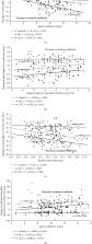
          Orthokeratology (OK) is widely used to slow the progression of myopia. Low-level laser therapy (LLLT) provides sufficient low energy to change the cellular function. This research is aimed at verifying the hypothesis that LLLT treatment could control myopia progression and comparing the abilities of OK lenses and LLLT to control the refractive error of myopia. Eighty-one children (81 eyes) who wore OK lenses, 74 children (74 eyes) who underwent LLLT treatment, and 74 children (74 eyes) who wore single-vision distance spectacles for 6 months were included. Changes in axial length (AL) were 0.23 ± 0.06 mm for children wearing spectacles, 0.06 ± 0.15 mm for children wearing OK lens, and −0.06 ± 0.15 mm for children treated with LLLT for 6 months. Changes in subfoveal choroidal thickness (SFChT) observed at the 6-month examination were −16.84 ± 7.85  μm, 14.98 ± 22.50  μm, and 35.30 ± 31.75  μm for the control group, OK group, and LLLT group, respectively. Increases in AL at 1 month and 6 months were significantly associated with age at LLLT treatment. Changes in AL were significantly correlated with the baseline spherical equivalent refraction (SER) and baseline AL in the OK and LLLT groups. Increases in SFChT at 1 month and 6 months were positively associated with age at enrolment for children wearing OK lens. At 6 months, axial elongation had decelerated in OK lens-wearers and LLLT-treated children. Slightly better myopia control was observed with LLLT treatment than with overnight OK lens-wearing. Evaluations of age, SER, and AL can enhance screening for high-risk myopia, improve the myopia prognosis, and help determine suitable control methods yielding the most benefits.

          Related collections

          Most cited references57

          • Record: found
          • Abstract: found
          • Article: not found

          The multifunctional choroid.

          The choroid of the eye is primarily a vascular structure supplying the outer retina. It has several unusual features: It contains large membrane-lined lacunae, which, at least in birds, function as part of the lymphatic drainage of the eye and which can change their volume dramatically, thereby changing the thickness of the choroid as much as four-fold over a few days (much less in primates). It contains non-vascular smooth muscle cells, especially behind the fovea, the contraction of which may thin the choroid, thereby opposing the thickening caused by expansion of the lacunae. It has intrinsic choroidal neurons, also mostly behind the central retina, which may control these muscles and may modulate choroidal blood flow as well. These neurons receive sympathetic, parasympathetic and nitrergic innervation. The choroid has several functions: Its vasculature is the major supply for the outer retina; impairment of the flow of oxygen from choroid to retina may cause Age-Related Macular Degeneration. The choroidal blood flow, which is as great as in any other organ, may also cool and warm the retina. In addition to its vascular functions, the choroid contains secretory cells, probably involved in modulation of vascularization and in growth of the sclera. Finally, the dramatic changes in choroidal thickness move the retina forward and back, bringing the photoreceptors into the plane of focus, a function demonstrated by the thinning of the choroid that occurs when the focal plane is moved back by the wearing of negative lenses, and, conversely, by the thickening that occurs when positive lenses are worn. In addition to focusing the eye, more slowly than accommodation and more quickly than emmetropization, we argue that the choroidal thickness changes also are correlated with changes in the growth of the sclera, and hence of the eye. Because transient increases in choroidal thickness are followed by a prolonged decrease in synthesis of extracellular matrix molecules and a slowing of ocular elongation, and attempts to decouple the choroidal and scleral changes have largely failed, it seems that the thickening of the choroid may be mechanistically linked to the scleral synthesis of macromolecules, and thus may play an important role in the homeostatic control of eye growth, and, consequently, in the etiology of myopia and hyperopia. Copyright 2009 Elsevier Ltd. All rights reserved.
            Bookmark
            • Record: found
            • Abstract: found
            • Article: not found

            The nuts and bolts of low-level laser (light) therapy.

            Soon after the discovery of lasers in the 1960s it was realized that laser therapy had the potential to improve wound healing and reduce pain, inflammation and swelling. In recent years the field sometimes known as photobiomodulation has broadened to include light-emitting diodes and other light sources, and the range of wavelengths used now includes many in the red and near infrared. The term "low level laser therapy" or LLLT has become widely recognized and implies the existence of the biphasic dose response or the Arndt-Schulz curve. This review will cover the mechanisms of action of LLLT at a cellular and at a tissular level and will summarize the various light sources and principles of dosimetry that are employed in clinical practice. The range of diseases, injuries, and conditions that can be benefited by LLLT will be summarized with an emphasis on those that have reported randomized controlled clinical trials. Serious life-threatening diseases such as stroke, heart attack, spinal cord injury, and traumatic brain injury may soon be amenable to LLLT therapy.
              Bookmark
              • Record: found
              • Abstract: found
              • Article: not found

              Retardation of myopia in Orthokeratology (ROMIO) study: a 2-year randomized clinical trial.

              This single-masked randomized clinical trial aimed to evaluate the effectiveness of orthokeratology (ortho-k) for myopic control. A total of 102 eligible subjects, ranging in age from 6 to 10 years, with myopia between 0.50 and 4.00 diopters (D) and astigmatism not more than 1.25D, were randomly assigned to wear ortho-k lenses or single-vision glasses for a period of 2 years. Axial length was measured by intraocular lens calculation by a masked examiner and was performed at the baseline and every 6 months. This study was registered at ClinicalTrials.gov, number NCT00962208. In all, 78 subjects (37 in ortho-k group and 41 in control group) completed the study. The average axial elongation, at the end of 2 years, were 0.36 ± 0.24 and 0.63 ± 0.26 mm in the ortho-k and control groups, respectively, and were significantly slower in the ortho-k group (P 0.54) but was correlated with the initial age of the subjects (P 1.00D per year) were 65% and 13% in younger (age range: 7-8 years) and older (age range: 9-10 years) children, respectively, in the control group and were 20% and 9%, respectively, in the ortho-k group. Five subjects discontinued ortho-k treatment due to adverse events. On average, subjects wearing ortho-k lenses had a slower increase in axial elongation by 43% compared with that of subjects wearing single-vision glasses. Younger children tended to have faster axial elongation and may benefit from early ortho-k treatment. (ClinicalTrials.gov number, NCT00962208.).
                Bookmark

                Author and article information

                Contributors
                Journal
                Biomed Res Int
                Biomed Res Int
                BMRI
                BioMed Research International
                Hindawi
                2314-6133
                2314-6141
                2021
                27 January 2021
                : 2021
                : 8915867
                Affiliations
                Affiliated Eye Hospital, Nanchang University, Nanchang 330006, China
                Author notes

                Academic Editor: Pietro Emanuele Napoli

                Author information
                https://orcid.org/0000-0002-1775-0655
                https://orcid.org/0000-0002-8334-9878
                https://orcid.org/0000-0003-1955-6570
                https://orcid.org/0000-0001-5254-9394
                https://orcid.org/0000-0002-9218-2408
                https://orcid.org/0000-0003-1408-1561
                https://orcid.org/0000-0003-4519-5051
                https://orcid.org/0000-0002-2469-4313
                https://orcid.org/0000-0001-5004-0498
                https://orcid.org/0000-0001-8572-6115
                https://orcid.org/0000-0003-3883-7730
                https://orcid.org/0000-0001-6004-8709
                Article
                10.1155/2021/8915867
                7861936
                33575355
                375f28ff-9d14-4024-9450-1311b4c6fbd9
                Copyright © 2021 Fen Xiong et al.

                This is an open access article distributed under the Creative Commons Attribution License, which permits unrestricted use, distribution, and reproduction in any medium, provided the original work is properly cited.

                History
                : 16 July 2020
                : 2 November 2020
                : 15 January 2021
                Funding
                Funded by: Science and Technology Program of Health Commission of Jiangxi Province
                Award ID: 202130555
                Categories
                Research Article

                Comments

                Comment on this article