6
views
0
recommends
+1 Recommend
0 collections
    0
    shares
      • Record: found
      • Abstract: found
      • Article: found
      Is Open Access

      Gastrointestinal ultrasonographic findings in cats with Feline panleukopenia: a case series

      research-article

      Read this article at

      Bookmark
          There is no author summary for this article yet. Authors can add summaries to their articles on ScienceOpen to make them more accessible to a non-specialist audience.

          Abstract

          Background

          Feline panleukopenia virus (FPV) is very resistant and highly contagious and infects domestic cats and other felids. FPV is particularly widespread among sheltered cats, and is associated with high morbidity and mortality, causing severe gastroenteritis characterized by anorexia, lethargy, fever, dehydration, hemorrhagic diarrhea, and vomiting. There is currently no data on the ultrasonographic features of cats affected with FPV. This case series describes abdominal ultrasonographic findings in shelter cats with naturally-occurring FPV, and assesses whether are associated with clinical and laboratory findings. Cats affected by FPV were enrolled in the study if an abdominal ultrasound was performed within 12 hours of diagnosis. Clinical, laboratory and survival data were collected from medical records. Ultrasonographic examinations were reviewed for gastrointestinal abnormalities and their associations with the above data were explored.

          Results

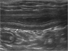
          Twenty-one cats were included. Nine cats (42.9%) died and 12 (57.1%) recovered. Based on ultrasonography, the duodenum and jejunum showed thinning of the mucosal layer in 70.6% and 66.6% of cats, thickening of the muscular layer in 52.9% and 57.1% of cats, and hyperechogenicity of the mucosa in 41.2% and 33.3%. Jejunal hyperechoic mucosal band paralleling the submucosa and irregular luminal surface were both observed in 33.3% of the cats. Survival was positively associated with increased jejunal mucosal echogenicity ( P = 0.003) and hyperechoic mucosal band ( P = 0.003). Peritoneal free fluid was positively associated with vomiting ( P = 0.002).

          Conclusions

          This study provides ultrasonographic features of naturally-occurring FPV in cats, which, as expected, are compatible with gastroenteropathy. The most frequent findings were diffuse small intestine mucosal layer thinning, muscular layer thickening and mucosal hyperechogenicity, jejunal hyperechoic mucosal band and irregular luminal surface. Ultrasonographic features may be useful to complete the clinical picture and assess the severity of the gastroenteropathy in FPV cats. Prospective studies are needed to confirm ultrasonographic prognostic factors.

          Related collections

          Most cited references22

          • Record: found
          • Abstract: found
          • Article: not found

          Adjusting for multiple testing--when and how?

          Multiplicity of data, hypotheses, and analyses is a common problem in biomedical and epidemiological research. Multiple testing theory provides a framework for defining and controlling appropriate error rates in order to protect against wrong conclusions. However, the corresponding multiple test procedures are underutilized in biomedical and epidemiological research. In this article, the existing multiple test procedures are summarized for the most important multiplicity situations. It is emphasized that adjustments for multiple testing are required in confirmatory studies whenever results from multiple tests have to be combined in one final conclusion and decision. In case of multiple significance tests a note on the error rate that will be controlled for is desirable.
            Bookmark
            • Record: found
            • Abstract: found
            • Article: not found

            Feline parvovirus infection and associated diseases.

            Feline panleukopenia, caused by the single-stranded DNA virus feline parvovirus (FPV), is a highly contagious and often lethal disease of cats and other Felidae. FPV, but also canine parvovirus (CPV) can be isolated from both healthy and diseased cats. In Germany, CPV was detected in only approximately 10% of feline samples, but in Southeast Asia, reports estimated that up to approximately 80% of diseased cats were infected with CPV. Infection spreads rapidly, especially in cells with high mitotic activity, such as bone marrow, lymphoid tissue and intestinal crypt cells. Anorexia, vomiting, diarrhoea, neutropenia and lymphopenia are common in clinically affected cases. In utero or neonatal infection can result in cerebellar hypoplasia. Depending on the severity of clinical signs, mortality ranges from 25 to 100%. Effective vaccination and thorough disinfection are of the utmost importance in the prevention of disease transmission in multi-cat households and animal shelters. If clinical signs develop, supportive treatment should be commenced. The efficacy of feline recombinant interferon and FPV antibodies has not been clearly demonstrated. Commercially available vaccines should induce protective immunity when administered according to current guidelines. Recent studies suggest that in some kittens, maternally derived antibodies (MDA) can persist for much longer than has been previously recognised. FPV serum antibody tests are available, but protection status needs to be interpreted with caution in kittens with MDA and a negative titre in adult cats does not necessarily denote lack of protection.
              Bookmark
              • Record: found
              • Abstract: found
              • Article: found
              Is Open Access

              Canine parvoviral enteritis: an update on the clinical diagnosis, treatment, and prevention

              Canine parvovirus type 2 is the cause of a highly contagious acute enteritis associated with high morbidity and mortality, with very low survival rates in untreated dogs. Although severe clinical disease typically occurs in dogs younger than 6 months of age, adults with insufficient immunity may potentially be affected. In this article, the current state of knowledge is reviewed regarding the diagnostic aspects of parvoviral enteritis, with special emphasis placed on the clinical relevance of the detection of viral antigens in the feces, detection of viral antibodies in the serum, or the polymerase chain reaction-based amplification of the viral DNA in the feces. In addition, the components of the supportive and symptomatic treatment aiming to optimize the outcome of the disease in the clinical setting are thoroughly reviewed. Immunization guidelines for the prevention of the disease are also updated.
                Bookmark

                Author and article information

                Contributors
                saraisaya1@gmail.com
                Journal
                BMC Vet Res
                BMC Vet Res
                BMC Veterinary Research
                BioMed Central (London )
                1746-6148
                7 January 2021
                7 January 2021
                2021
                : 17
                : 20
                Affiliations
                [1 ]Istituto Veterinario di Novara, 28060 Granozzo con Monticello (NO), Italy
                [2 ]GRID grid.7644.1, ISNI 0000 0001 0120 3326, Department of Veterinary Medicine, , University of Bari “Aldo Moro”, ; BA 70010 Valenzano, Italy
                [3 ]GRID grid.5608.b, ISNI 0000 0004 1757 3470, Department of Animal Medicine, Production and Health, , University of Padova, ; Legnaro, PD Italy
                [4 ]GRID grid.7400.3, ISNI 0000 0004 1937 0650, Clinic for Small Animal Internal Medicine, Vetsuisse Faculty, , University of Zurich, ; Winterthurerstrasse 260, 8057 Zurich, Switzerland
                Article
                2720
                10.1186/s12917-020-02720-w
                7791690
                33413382
                36f17e54-59f4-4c42-9a94-a9f0d0c32659
                © The Author(s) 2021

                Open AccessThis article is licensed under a Creative Commons Attribution 4.0 International License, which permits use, sharing, adaptation, distribution and reproduction in any medium or format, as long as you give appropriate credit to the original author(s) and the source, provide a link to the Creative Commons licence, and indicate if changes were made. The images or other third party material in this article are included in the article's Creative Commons licence, unless indicated otherwise in a credit line to the material. If material is not included in the article's Creative Commons licence and your intended use is not permitted by statutory regulation or exceeds the permitted use, you will need to obtain permission directly from the copyright holder. To view a copy of this licence, visit http://creativecommons.org/licenses/by/4.0/. The Creative Commons Public Domain Dedication waiver ( http://creativecommons.org/publicdomain/zero/1.0/) applies to the data made available in this article, unless otherwise stated in a credit line to the data.

                History
                : 13 March 2020
                : 9 December 2020
                Funding
                Funded by: FundRef http://dx.doi.org/10.13039/501100003500, Università degli Studi di Padova;
                Award ID: BIRD168451
                Categories
                Research Article
                Custom metadata
                © The Author(s) 2021

                Veterinary medicine
                ultrasound,fpv,cat,gastrointestinal tract
                Veterinary medicine
                ultrasound, fpv, cat, gastrointestinal tract

                Comments

                Comment on this article