49
views
0
recommends
+1 Recommend
0 collections
    4
    shares
      • Record: found
      • Abstract: found
      • Article: found
      Is Open Access

      Steady-State Visual Evoked Potentials Can Be Explained by Temporal Superposition of Transient Event-Related Responses

      research-article

      Read this article at

      Bookmark
          There is no author summary for this article yet. Authors can add summaries to their articles on ScienceOpen to make them more accessible to a non-specialist audience.

          Abstract

          Background

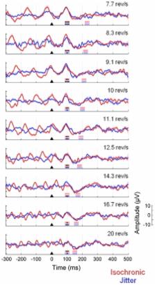
          One common criterion for classifying electrophysiological brain responses is based on the distinction between transient (i.e. event-related potentials, ERPs) and steady-state responses (SSRs). The generation of SSRs is usually attributed to the entrainment of a neural rhythm driven by the stimulus train. However, a more parsimonious account suggests that SSRs might result from the linear addition of the transient responses elicited by each stimulus. This study aimed to investigate this possibility.

          Methodology/Principal Findings

          We recorded brain potentials elicited by a checkerboard stimulus reversing at different rates. We modeled SSRs by sequentially shifting and linearly adding rate-specific ERPs. Our results show a strong resemblance between recorded and synthetic SSRs, supporting the superposition hypothesis. Furthermore, we did not find evidence of entrainment of a neural oscillation at the stimulation frequency.

          Conclusions/Significance

          This study provides evidence that visual SSRs can be explained as a superposition of transient ERPs. These findings have critical implications in our current understanding of brain oscillations. Contrary to the idea that neural networks can be tuned to a wide range of frequencies, our findings rather suggest that the oscillatory response of a given neural network is constrained within its natural frequency range.

          Related collections

          Most cited references49

          • Record: found
          • Abstract: found
          • Article: not found

          Mining event-related brain dynamics.

          This article provides a new, more comprehensive view of event-related brain dynamics founded on an information-based approach to modeling electroencephalographic (EEG) dynamics. Most EEG research focuses either on peaks 'evoked' in average event-related potentials (ERPs) or on changes 'induced' in the EEG power spectrum by experimental events. Although these measures are nearly complementary, they do not fully model the event-related dynamics in the data, and cannot isolate the signals of the contributing cortical areas. We propose that many ERPs and other EEG features are better viewed as time/frequency perturbations of underlying field potential processes. The new approach combines independent component analysis (ICA), time/frequency analysis, and trial-by-trial visualization that measures EEG source dynamics without requiring an explicit head model.
            Bookmark
            • Record: found
            • Abstract: found
            • Article: not found

            Human EEG responses to 1-100 Hz flicker: resonance phenomena in visual cortex and their potential correlation to cognitive phenomena.

            The individual properties of visual objects, like form or color, are represented in different areas in our visual cortex. In order to perceive one coherent object, its features have to be bound together. This was found to be achieved in cat and monkey brains by temporal correlation of the firing rates of neurons which code the same object. This firing rate is predominantly observed in the gamma frequency range (approx. 30-80 Hz, mainly around 40 Hz). In addition, it has been shown in humans that stimuli which flicker at gamma frequencies are processed faster by our brains than when they flicker at different frequencies. These effects could be due to neural oscillators, which preferably oscillate at certain frequencies, so-called resonance frequencies. It is also known that neurons in visual cortex respond to flickering stimuli at the frequency of the flickering light. If neural oscillators exist with resonance frequencies, they should respond more strongly to stimulation with their resonance frequency. We performed an experiment, where ten human subjects were presented flickering light at frequencies from 1 to 100 Hz in 1-Hz steps. The event-related potentials exhibited steady-state oscillations at all frequencies up to at least 90 Hz. Interestingly, the steady-state potentials exhibited clear resonance phenomena around 10, 20, 40 and 80 Hz. This could be a potential neural basis for gamma oscillations in binding experiments. The pattern of results resembles that of multiunit activity and local field potentials in cat visual cortex.
              Bookmark
              • Record: found
              • Abstract: found
              • Article: not found

              Natural frequencies of human corticothalamic circuits.

              The frequency tuning of a system can be directly determined by perturbing it and by observing the rate of the ensuing oscillations, the so called natural frequency. This approach is used, for example, in physics, in geology, and also when one tunes a musical instrument. In the present study, we employ transcranial magnetic stimulation (TMS) to directly perturb a set of selected corticothalamic modules (Brodmann areas 19, 7, and 6) and high-density electroencephalogram to measure their natural frequency. TMS consistently evoked dominant alpha-band oscillations (8-12 Hz) in the occipital cortex, beta-band oscillations (13-20 Hz) in the parietal cortex, and fast beta/gamma-band oscillations (21-50 Hz) in the frontal cortex. Each cortical area tended to preserve its own natural frequency also when indirectly engaged by TMS through brain connections and when stimulated at different intensities, indicating that the observed oscillations reflect local physiological mechanisms. These findings were reproducible across individuals and represent the first direct characterization of the coarse electrophysiological properties of three associative areas of the human cerebral cortex. Most importantly, they indicate that, in healthy subjects, each corticothalamic module is normally tuned to oscillate at a characteristic rate. The natural frequency can be directly measured in virtually any area of the cerebral cortex and may represent a straightforward and flexible way to probe the state of human thalamocortical circuits at the patient's bedside.
                Bookmark

                Author and article information

                Contributors
                Role: Editor
                Journal
                PLoS One
                plos
                plosone
                PLoS ONE
                Public Library of Science (San Francisco, USA )
                1932-6203
                2011
                18 January 2011
                : 6
                : 1
                : e14543
                Affiliations
                [1 ]Centre for Cognitive Neuroimaging (CCNi), Institute for Neuroscience and Psychology, University of Glasgow, Glasgow, United Kingdom
                [2 ]Department of Biological and Health Psychology, Autonoma University of Madrid, Madrid, Spain
                [3 ]Department of Clinical Psychology and Psychobiology, University of Santiago de Compostela, Santiago de Compostela, Spain
                [4 ]Center of Magnetoencephalography Dr Perez Modrego, Complutense University of Madrid, Madrid, Spain
                [5 ]Laboratory of Cognitive and Computational Neuroscience, Center for Biomedical Technology, Complutense University of Madrid-Polytechnic University of Madrid, Madrid, Spain
                [6 ]Department of Basic Psychology, Autonoma University of Madrid, Madrid, Spain
                [7 ]School of Psychology, University of Glasgow, Glasgow, United Kingdom
                Cuban Neuroscience Center, Cuba
                Author notes

                Conceived and designed the experiments: AC PPA. Performed the experiments: AC AD PC. Analyzed the data: AC PPA JG. Wrote the paper: AC PPA AD PC JG.

                Article
                10-PONE-RA-20101R2
                10.1371/journal.pone.0014543
                3022588
                21267081
                36a1e74f-c73e-4a4d-852b-8f881d1abd42
                Capilla et al. This is an open-access article distributed under the terms of the Creative Commons Attribution License, which permits unrestricted use, distribution, and reproduction in any medium, provided the original author and source are credited.
                History
                : 22 June 2010
                : 10 December 2010
                Page count
                Pages: 15
                Categories
                Research Article
                Neuroscience/Cognitive Neuroscience
                Neuroscience/Sensory Systems
                Neuroscience/Theoretical Neuroscience

                Uncategorized
                Uncategorized

                Comments

                Comment on this article