14
views
0
recommends
+1 Recommend
0 collections
    0
    shares
      • Record: found
      • Abstract: found
      • Article: found
      Is Open Access

      Alteration of fatty acid oxidation by increased CPT1A on replicative senescence of placenta-derived mesenchymal stem cells

      research-article

      Read this article at

      Bookmark
          There is no author summary for this article yet. Authors can add summaries to their articles on ScienceOpen to make them more accessible to a non-specialist audience.

          Abstract

          Background

          Human placenta-derived mesenchymal stem cells (PD-MSCs) are powerful sources for cell therapy in regenerative medicine. However, a limited lifespan by senescence through mechanisms that are well unknown is the greatest obstacle. In the present study, we first demonstrated the characterization of replicative senescent PD-MSCs and their possible mitochondrial functional alterations.

          Methods

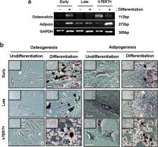
          Human PD-MSCs were cultured to senescent cells for a long period of time. The cells of before passage number 8 were early cells and after passage number 14 were late cells. Also, immortalized cells of PD-MSCs (overexpressed hTERT gene into PD-MSCs) after passage number 14 were positive control of non-senescent cells. The characterization and mitochondria analysis of PD-MSCs were explored with long-term cultivation.

          Results

          Long-term cultivation of PD-MSCs exhibited increases of senescent markers such as SA-β-gal and p21 including apoptotic factor, and decreases of proliferation, differentiation potential, and survival factor. Mitochondrial dysfunction was also observed in membrane potential and metabolic flexibility with enlarged mitochondrial mass. Interestingly, we founded that fatty acid oxidation (FAO) is an important metabolism in PD-MSCs, and carnitine palmitoyltransferase1A (CPT1A) overexpressed in senescent PD-MSCs. The inhibition of CPT1A induced a change of energy metabolism and reversed senescence of PD-MSCs.

          Conclusions

          These findings suggest that alteration of FAO by increased CPT1A plays an important role in mitochondrial dysfunction and senescence of PD-MSCs during long-term cultivation.

          Related collections

          Most cited references30

          • Record: found
          • Abstract: found
          • Article: found
          Is Open Access

          Mitochondrial effectors of cellular senescence: beyond the free radical theory of aging

          Cellular senescence is a process that results from a variety of stresses, leading to a state of irreversible growth arrest. Senescent cells accumulate during aging and have been implicated in promoting a variety of age-related diseases. Mitochondrial stress is an effective inducer of cellular senescence, but the mechanisms by which mitochondria regulate permanent cell growth arrest are largely unexplored. Here, we review some of the mitochondrial signaling pathways that participate in establishing cellular senescence. We discuss the role of mitochondrial reactive oxygen species (ROS), mitochondrial dynamics (fission and fusion), the electron transport chain (ETC), bioenergetic balance, redox state, metabolic signature, and calcium homeostasis in controlling cellular growth arrest. We emphasize that multiple mitochondrial signaling pathways, besides mitochondrial ROS, can induce cellular senescence. Together, these pathways provide a broader perspective for studying the contribution of mitochondrial stress to aging, linking mitochondrial dysfunction and aging through the process of cellular senescence.
            Bookmark
            • Record: found
            • Abstract: found
            • Article: not found

            Mitochondrial dysfunction contributes to oncogene-induced senescence.

            The expression of oncogenic ras in normal human cells quickly induces an aberrant proliferation response that later is curtailed by a cell cycle arrest known as cellular senescence. Here, we show that cells expressing oncogenic ras display an increase in the mitochondrial mass, the mitochondrial DNA, and the mitochondrial production of reactive oxygen species (ROS) prior to the senescent cell cycle arrest. By the time the cells entered senescence, dysfunctional mitochondria accumulated around the nucleus. The mitochondrial dysfunction was accompanied by oxidative DNA damage, a drop in ATP levels, and the activation of AMPK. The increase in mitochondrial mass and ROS in response to oncogenic ras depended on intact p53 and Rb tumor suppression pathways. In addition, direct interference with mitochondrial functions by inhibiting the expression of the Rieske iron sulfur protein of complex III or the use of pharmacological inhibitors of the electron transport chain and oxidative phosphorylation was sufficient to trigger senescence. Taking these results together, this work suggests that mitochondrial dysfunction is an effector pathway of oncogene-induced senescence.
              Bookmark
              • Record: found
              • Abstract: found
              • Article: not found

              Adiponectin increases fatty acid oxidation in skeletal muscle cells by sequential activation of AMP-activated protein kinase, p38 mitogen-activated protein kinase, and peroxisome proliferator-activated receptor alpha.

              Adiponectin has recently received a great deal of attention due to its beneficial effects on insulin resistance and metabolic disorders. One of the mechanisms through which adiponectin exerts such effects involves an increase in fatty acid oxidation in muscle and liver. In the present study, we demonstrate that 5'-AMP-activated protein kinase (AMPK) and p38 mitogen-activated protein kinase (MAPK) are involved in the activation of peroxisome proliferator-activated receptor (PPAR)alpha by adiponectin in muscle cells. Adiponectin increases the transcriptional activity of PPARalpha and the expression of its target genes, including ACO, CPT1, and FABP3 in C2C12 myotubes. These effects were suppressed by the overexpression of a dominant-negative form of AMPK. Moreover, chemical inhibitors of AMPK and p38 MAPK potently repressed fatty acid oxidation and the induction of PPARalpha target gene expression by adiponectin. Interestingly, araA, an AMPK inhibitor, prevented the activation of p38 MAPK, whereas SB203580, a p38 MAPK inhibitor, did not affect AMPK activation, suggesting that p38 MAPK is a downstream signaling factor of AMPK. Taken together, these results suggest that adiponectin stimulates fatty acid oxidation in muscle cells by the sequential activation of AMPK, p38 MAPK, and PPARalpha.
                Bookmark

                Author and article information

                Contributors
                gjkim@cha.ac.kr
                Journal
                Stem Cell Res Ther
                Stem Cell Res Ther
                Stem Cell Research & Therapy
                BioMed Central (London )
                1757-6512
                3 January 2020
                3 January 2020
                2020
                : 11
                : 1
                Affiliations
                [1 ]ISNI 0000 0004 0647 3511, GRID grid.410886.3, Department of Biomedical Science, , CHA University, ; 689, Sampyeong-dong, Bundang-gu, Seongnam-si, Gyeonggi-do Republic of Korea
                [2 ]ISNI 0000 0001 0840 2678, GRID grid.222754.4, Department of Anatomy, , Korea University College of Medicine, ; Seoul, Republic of Korea
                [3 ]ISNI 0000 0001 0842 2126, GRID grid.413967.e, Department of Pathology, , University of Ulsan College of Medicine, Asan Medical Center, ; Seoul, Republic of Korea
                Author information
                http://orcid.org/0000-0002-2320-7157
                Article
                1471
                10.1186/s13287-019-1471-y
                6941254
                31900237
                365ab2a1-6ee8-486c-b0fc-35d802184572
                © The Author(s). 2019

                Open AccessThis article is distributed under the terms of the Creative Commons Attribution 4.0 International License ( http://creativecommons.org/licenses/by/4.0/), which permits unrestricted use, distribution, and reproduction in any medium, provided you give appropriate credit to the original author(s) and the source, provide a link to the Creative Commons license, and indicate if changes were made. The Creative Commons Public Domain Dedication waiver ( http://creativecommons.org/publicdomain/zero/1.0/) applies to the data made available in this article, unless otherwise stated.

                History
                : 30 April 2019
                : 10 October 2019
                : 25 October 2019
                Funding
                Funded by: FundRef http://dx.doi.org/10.13039/501100003625, Ministry of Health and Welfare;
                Award ID: HI17C1050
                Award Recipient :
                Funded by: FundRef http://dx.doi.org/10.13039/501100003725, National Research Foundation of Korea;
                Award ID: NRF-2017M3A9B406
                Award Recipient :
                Categories
                Research
                Custom metadata
                © The Author(s) 2020

                Molecular medicine
                placenta-derived mesenchymal stem cell,senescence,mitochondria,fatty acid,cpt1a
                Molecular medicine
                placenta-derived mesenchymal stem cell, senescence, mitochondria, fatty acid, cpt1a

                Comments

                Comment on this article