17
views
0
recommends
+1 Recommend
0 collections
    0
    shares
      • Record: found
      • Abstract: found
      • Article: found
      Is Open Access

      Your Brain on Art: Emergent Cortical Dynamics During Aesthetic Experiences

      research-article

      Read this article at

      Bookmark
          There is no author summary for this article yet. Authors can add summaries to their articles on ScienceOpen to make them more accessible to a non-specialist audience.

          Abstract

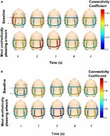
          The brain response to conceptual art was studied with mobile electroencephalography (EEG) to examine the neural basis of aesthetic experiences. In contrast to most studies of perceptual phenomena, participants were moving and thinking freely as they viewed the exhibit The Boundary of Life is Quietly Crossed by Dario Robleto at the Menil Collection-Houston. The brain activity of over 400 subjects was recorded using dry-electrode and one reference gel-based EEG systems over a period of 3 months. Here, we report initial findings based on the reference system. EEG segments corresponding to each art piece were grouped into one of three classes (complex, moderate, and baseline) based on analysis of a digital image of each piece. Time, frequency, and wavelet features extracted from EEG were used to classify patterns associated with viewing art, and ranked based on their relevance for classification. The maximum classification accuracy was 55% (chance = 33%) with delta and gamma features the most relevant for classification. Functional analysis revealed a significant increase in connection strength in localized brain networks while subjects viewed the most aesthetically pleasing art compared to viewing a blank wall. The direction of signal flow showed early recruitment of broad posterior areas followed by focal anterior activation. Significant differences in the strength of connections were also observed across age and gender. This work provides evidence that EEG, deployed on freely behaving subjects, can detect selective signal flow in neural networks, identify significant differences between subject groups, and report with greater-than-chance accuracy the complexity of a subject's visual percept of aesthetically pleasing art. Our approach, which allows acquisition of neural activity “in action and context,” could lead to understanding of how the brain integrates sensory input and its ongoing internal state to produce the phenomenon which we term aesthetic experience.

          Related collections

          Most cited references48

          • Record: found
          • Abstract: not found
          • Article: not found

          Modulation of Oscillatory Neuronal Synchronization by Selective Visual Attention

          P. Fries (2001)
            Bookmark
            • Record: found
            • Abstract: found
            • Article: not found

            Cross-frequency phase-phase coupling between θ and γ oscillations in the hippocampus.

            Neuronal oscillations allow for temporal segmentation of neuronal spikes. Interdependent oscillators can integrate multiple layers of information. We examined phase-phase coupling of theta and gamma oscillators in the CA1 region of rat hippocampus during maze exploration and rapid eye movement sleep. Hippocampal theta waves were asymmetric, and estimation of the spatial position of the animal was improved by identifying the waveform-based phase of spiking, compared to traditional methods used for phase estimation. Using the waveform-based theta phase, three distinct gamma bands were identified: slow gamma(S) (gamma(S); 30-50 Hz), midfrequency gamma(M) (gamma(M); 50-90 Hz), and fast gamma(F) (gamma(F); 90-150 Hz or epsilon band). The amplitude of each sub-band was modulated by the theta phase. In addition, we found reliable phase-phase coupling between theta and both gamma(S) and gamma(M) but not gamma(F) oscillators. We suggest that cross-frequency phase coupling can support multiple time-scale control of neuronal spikes within and across structures.
              Bookmark
              • Record: found
              • Abstract: found
              • Article: not found

              Orbitofrontal cortex and its contribution to decision-making.

              Damage to orbitofrontal cortex (OFC) produces an unusual pattern of deficits. Patients have intact cognitive abilities but are impaired in making everyday decisions. Here we review anatomical, neuropsychological, and neurophysiological evidence to determine the neuronal mechanisms that might underlie these impairments. We suggest that OFC plays a key role in processing reward: It integrates multiple sources of information regarding the reward outcome to derive a value signal. In effect, OFC calculates how rewarding a reward is. This value signal can then be held in working memory where it can be used by lateral prefrontal cortex to plan and organize behavior toward obtaining the outcome, and by medial prefrontal cortex to evaluate the overall action in terms of its success and the effort that was required. Thus, acting together, these prefrontal areas can ensure that our behavior is most efficiently directed towards satisfying our needs.
                Bookmark

                Author and article information

                Contributors
                Journal
                Front Hum Neurosci
                Front Hum Neurosci
                Front. Hum. Neurosci.
                Frontiers in Human Neuroscience
                Frontiers Media S.A.
                1662-5161
                18 November 2015
                2015
                : 9
                : 626
                Affiliations
                [1] 1Office of Science and Engineering Laboratories, Division of Biomedical Physics, Center for Devices and Radiological Health, U.S. Food and Drug Administration Silver Spring, MD, USA
                [2] 2Laboratory for Non-Invasive Brain Machine Interfaces, Department of Electrical and Computer Engineering, University of Houston Houston, TX, USA
                [3] 3American Artist Houston, TX, USA
                [4] 4The Menil Collection Houston, TX, USA
                Author notes

                Edited by: Marcos Nadal, University of Vienna, Austria

                Reviewed by: Juliana Yordanova, Bulgarian Academy of Sciences, Bulgaria; Daniela M. Pfabigan, University of Vienna, Austria; Stephen Grossberg, Boston University, USA

                *Correspondence: Kimberly L. Kontson kimberly.kontson@ 123456fda.hhs.gov

                †These authors have contributed equally to this work.

                Article
                10.3389/fnhum.2015.00626
                4649259
                25653611
                3542d76c-1427-4f4b-9919-69bea3f84660
                Copyright © 2015 Kontson, Megjhani, Brantley, Cruz-Garza, Nakagome, Robleto, White, Civillico and Contreras-Vidal.

                This is an open-access article distributed under the terms of the Creative Commons Attribution License (CC BY). The use, distribution or reproduction in other forums is permitted, provided the original author(s) or licensor are credited and that the original publication in this journal is cited, in accordance with accepted academic practice. No use, distribution or reproduction is permitted which does not comply with these terms.

                History
                : 15 June 2015
                : 02 November 2015
                Page count
                Figures: 13, Tables: 1, Equations: 6, References: 69, Pages: 17, Words: 11051
                Categories
                Neuroscience
                Original Research

                Neurosciences
                eeg,machine learning,functional connectivity (fc),aesthetics,freely moving
                Neurosciences
                eeg, machine learning, functional connectivity (fc), aesthetics, freely moving

                Comments

                Comment on this article